生命科学研究

生命科学研究

生命科学研究

在生命科学研究中心,您可以掌握最新的关于先进显微镜、成像技术、电镜样品制备和图像分析的前沿应用和创新,涵盖的主题包括细胞生物学、神经科学和癌症研究。希望在这里可以帮助您提升研究能力和精进显微镜在各个科学领域实际应用,并了解徕卡如何通过精确的可视化、图像解读和推进研究进展来赋能您的工作。
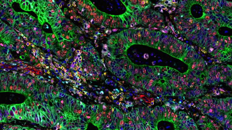
Cell DIVE multiplexed image of FFPE tissue section from human colon adenocarcinoma tissue.

Multiplexed Imaging Reveals Tumor Immune Landscape in Colon Cancer

Cancer immunotherapy benefits few due to resistance and relapse, and combinatorial therapeutic strategies that target multiple steps of the cancer-immunity cycle may improve outcomes. This study shows…
Cell DIVE multiplexed image of FFPE tissue section from human invasive ductal carcinoma (IDC)

AI-Powered Hi-Plex Spatial Analysis Tools for Breast Cancer Research

Breast cancer (BC) is the leading cause of cancer-related deaths in women. Investigating the tumor microenvironment (TME) is crucial to elucidate the mechanisms of tumor progression. Systematic…
Zebrafish-embryo image captured using a THUNDER Imager Tissue and live instant computational clearing.

Improving Zebrafish-Embryo Screening with Fast, High-Contrast Imaging

Discover from this article how screening of transgenic zebrafish embryos is boosted with high-speed, high-contrast imaging using the DM6 B microscope, ensuring accurate targeting for developmental…
Cell DIVE multiplexed image of FFPE tissue section from syngeneic murine cancer model, 4T1.

利用人工智能驱动的空间蛋白质组学绘制肿瘤免疫图谱

未经治疗肿瘤的空间图谱分析可呈现肿瘤免疫结构的整体特征,有助于理解治疗反应。具有免疫活性的小鼠模型对于识别肿瘤发生发展过程中免疫依赖性事件至关重要。要表征这些具有完整免疫系统及相互作用细胞组分的模型,需要采用多重标记分析技术。我们展示了一种基于人工智能的空间蛋白质组学方法,用于研究小鼠癌组织中的肿瘤-免疫互作机制。
Automated Laser Microdissection for Proteome Analysis

深度视觉蛋白质组学提供精确的空间蛋白质组信息

尽管可使用基于成像和质谱的方法进行空间蛋白质组学研究,但是图像与单细胞分辨率蛋白丰度测量值的关联仍然是个巨大的挑战。最近引入的一种方法,深层视觉蛋白质组学(DVP),将细胞表型的人工智能图像分析与自动化的单细胞或单核激光显微切割及超高灵敏度的质谱分析结合在了一起。DVP在保留空间背景的同时,将蛋白丰度与复杂的细胞或亚细胞表型关联在一起。
Multiplexed Cell DIVE imaging of Adult Human Alzheimer’s brain tissue section demonstrating expression of markers specific to astrocytes (GFAP, S100B), microglia (TMEM119, IBA1), AD-associated markers (p-Tau217, β-amyloid) and immune cells such as CD11b+, CD163+, CD4+, and HLA-DRA+, clustered around the β-amyloid plaques.

阿尔茨海默病神经免疫相互作用的空间分析

阿尔茨海默病(AD)是一种复杂的神经退行性疾病,以神经原纤维缠结、β-淀粉样斑块和神经炎症为特征。这些功能障碍由局部免疫反应触发或加剧。因此,在空间背景下理解神经免疫相互作用对于阐明 AD 发病机制至关重要。本研究采用 Cell DIVE 多重成像技术和 Aivia 人工智能辅助空间分析工具,探究 AD 病理标志物周围免疫细胞的特征。
Intestinal organoids label with FUCCI reporter to follow cell cycle dynamics. Courtesy of Franziska Moos. Liberali lab. FMI Basel (Switzerland).

双视野光片显微镜,适用于大型多细胞系统

展示复杂多细胞系统的动态是生物学中的一个基本目标。为了应对在大型时空尺度上进行活体成像的挑战,作者在《自然·方法》杂志上发表的一篇论文中介绍了一种开放式多样本双视野光片显微镜。研究发现,Viventis LS2 Live显微镜在以单细胞分辨率成像大型样本方面取得了显著进展。

肿瘤空间微环境的元癌症分析

研究 TME中肿瘤、基质和免疫细胞之间的相互作用需要采用超多重免疫荧光成像方法。在这里,我们分析了一组Cell Signaling Technology(CST®)抗体,这些抗体针对肺癌、结肠癌和胰腺癌等癌症的标志物。通过Cell DIVE成像和Aivia中的聚类分析,我们确定了TME中的空间相互作用,包括组织特异性和共有的相互作用。
Multiplexed Cell DIVE imaging of Colon Adenocarcinoma (CAC) tissue. A panel of approximately 30 biomarkers targeted towards various leukocyte lineages, epithelial, stromal, and endothelial cell types was utilized to characterize the tumor immune microenvironment in human colon adenocarcinoma (CAC) tissue.

通过成像和AI绘制结直肠癌的景观

结肠癌是一种高负担疾病。尽管进行了化疗干预和手术切除,但疾病可能会复发。了解结肠癌微环境对于改善治疗效果是必要的。在这里,我们使用空间生物学方法,通过Cell DIVE和 Aivia可视化结肠腺癌组织中的30个生物标志物。我们探讨了肿瘤组织的血管化、免疫细胞反应和细胞增殖。
Scroll to top