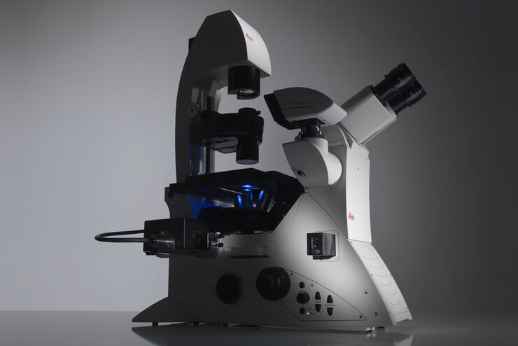
THUNDER Imager Cell Spinning Disk系统
倒置显微镜
复合光学显微镜
产品
首页
Leica Microsystems
THUNDER Imager Cell Spinning Disk系统
通过协同作用提高清晰度
阅读我们的最新文章
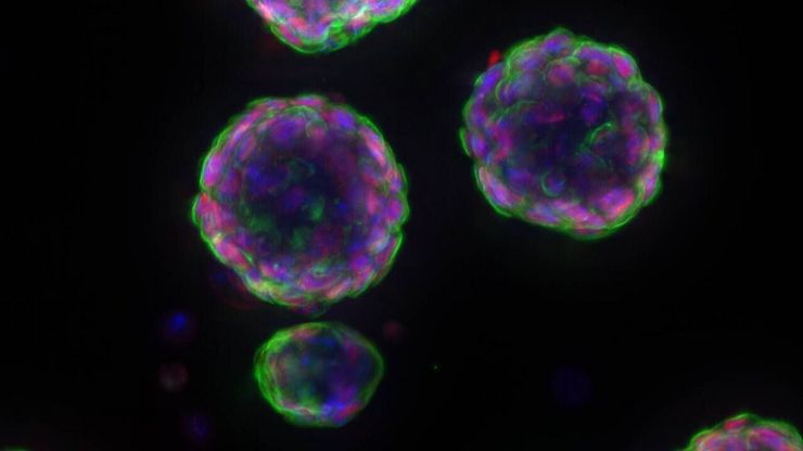
 and astrocytes (green) in a cortical spheroid derived from human induced pluripotent stem cells.")
活细胞成像指南
在生命科学各研究领域的广泛应用中,活细胞成像是一种不可或缺的工具,用于观察细胞在尽可能接近活体(即活的、活跃的)状态下的情况。本指南回顾了确保成功进行活细胞成像的各种重要注意事项,并介绍了各种旨在克服常见挑战的高性能解决方案。这些进展使我们能够对细胞生理学和动力学有新的认识。
Factors to Consider When Selecting a Research Microscope
An optical microscope is often one of the central devices in a life-science research lab. It can be used for various applications which shed light on many scientific questions. Thereby the…
 and tubulin (magenta), acquired using Viventis Deep. Courtesy of Akanksha Jain, Treutlein Lab ETH-DBSSE Basel (Switzerland).")
如何深入了解类器官和细胞球模型
在本电子书中,您将了解3D细胞培养模型(如类器官和细胞球)成像的关键注意事项。探索创新型显微镜解决方案,来实时记录类器官和细胞球的动态成像过程。

线虫研究指南 - 针对线虫的相关工作
本指南概述了可以高效进行线虫的研究显微镜技术。线虫是一种广泛使用的模式生物,与人类有大约 70% 的基因同源性,是研究发育、神经科学、遗传学和衰老的理想生物。它的透明性和易培育性使其成为一个出色的遗传学模型系统。它可以进行高分辨率成像。主要的实验方法包括挑虫、转基因、荧光筛选、成像和记录。

Boston and San Francisco Innovation Hubs
Boston and San Francisco Innovation Hubs are here to help you advance scientific discovery. We provide researchers access to state-of-the-art microscope technology and expert guidance. Located in the…

揭开类器官模型在生物医学研究中的秘密
准备深入了解类器官和3D培养物的世界,它们是促进我们了解人类健康的重要工具。浏览这些复杂的结构并获取清晰的图像进行分析是一项挑战。在本次活动中,来自牛津大学和伦敦大学学院的研究人员将与我们一起展示Thunder Imager Cell转盘共聚焦系统 如何提供更有说服力的高质量数据,以便深入了解各种模型。

全内反射荧光显微镜(total internal reflection fluorescent microscope,TIRFM)在生命科学研究中的应用
全内反射荧光显微镜的独特之处在于利用衰逝波激发荧光团。与传统的弧光灯、LED 或激光宽场荧光照明方式不同,衰逝波仅能从盖玻片/介质界面开始穿透样本约 100 纳米深度。

全内反射荧光(total internal reflection fluorescent microscope,TIRF)显微镜
全内反射荧光(TIRF)是荧光显微镜技术中的一项特殊技术,由密歇根大学安娜堡分校的 Daniel Axelrod 于 1980 年代初开发。TIRF 显微镜能提供轴向分辨率低于 100 纳米的超高清晰图像,这使得观察膜相关过程成为可能。
生物制药
对于生物制药行业,Leica 解决方案有助于加快药物发现,增强细胞分析,并支持符合法规的数据完整性。

Advanced Tissue Imaging & Analysis
Gain insights into tissue structure and function to improve your understanding of spatial biology and disease mechanisms with advanced imaging solutions from Leica Microsystems.
应用领域
类器官和3D细胞培养
生命科学研究中最令人振奋的最新进展之一是3D细胞培养系统的发展,例如类器官、球状体或器官芯片模型。 3D细胞培养物是一种人工环境,在这种环境中,细胞能够在三维空间中生长并与周围环境相互作用。 这些环境条件与它们在体内的情况相似。
生物制药
对于生物制药行业,Leica 解决方案有助于加快药物发现,增强细胞分析,并支持符合法规的数据完整性。
