STELLARIS CRS
共聚焦显微镜
产品
首页
Leica Microsystems
STELLARIS CRS 相干拉曼散射显微镜
了解无标记化学显微成像
阅读我们的最新文章
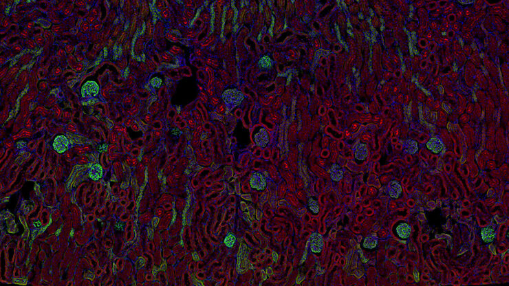
 dataset, showing the biochemically distinct structures of a fresh, untreated apple slice.")
如何为受激拉曼散射(SRS)成像制备样品
请参阅以下关于受激拉曼散射(SRS)的样本制备、图像采集、数据分析和流程开发的指南。受激拉曼散射光谱成像技术也被称为SRS显微技术。
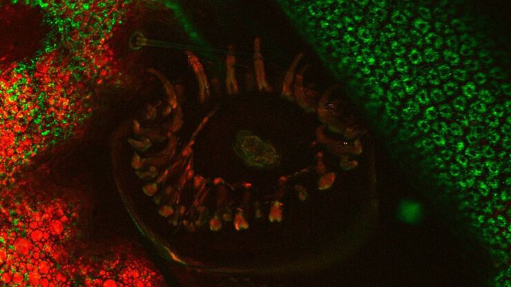
, unsaturated lipids (magenta, 3050 cm-1), collagen (SHG, cyan). Sample courtesy of R. Rudolf, J Klicks, Hochschule Mannheim")
相干拉曼散射显微镜的潜力一瞥
相干拉曼散射显微镜(CRS)是一种强大的无标记化学特异性成像方法。它基于样品中分子的固有振动对比特征。
CRS 可提供有关细胞、组织和完整模式生物体内生化组成和代谢过程的高分辨率(亚细胞水平)和动态(高达视频速率)信息。它还能在不干扰小分子功能的情况下对其进行成像。这些信息与荧光显微镜提供的分子对比具有高度协同作用。毫不奇怪,CRS…

用SRS显微镜对配方产品进行表征分析
从药品和消费者健康产品到农用化学品和油漆,霜剂、糊剂、凝胶、乳剂和片剂常见于众多制造领域。为提高有效性以及产品性能和安全性,有必要了解产品中各成分之间的相互作用。具备能评估活性成分的结构、稳定性并对其输送进行可视化的技术对配方产品制造业而言具有重大价值。

模式生物研究
模式生物是研究人员用来研究特定生物学过程的物种。 它们具有与人类相似的遗传特征,通常用于遗传学、发育生物学和神经科学等研究领域。 选择模式生物的原因通常是它们在实验室环境中易于保持和繁殖、生成周期短,或能够产生突变体来研究某些性状或疾病。
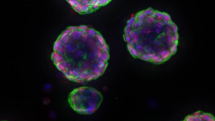
癌症研究
癌症是一种复杂的异质性疾病,由于细胞生长失控而引起。 一个或一组细胞的基因和表观遗传的变化破坏了正常功能,导致细胞自发、不受控制地生长和增殖。
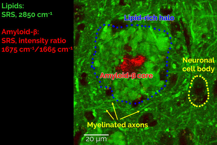
受激拉曼散射显微镜探测神经退行性疾病
Despite decades of research, the molecular mechanisms underlying some of the most severe neurodegenerative diseases, such as Alzheimer’s or Parkinson’s, remain poorly understood. The progression of…

CARS 相干反斯托克斯拉曼散射显微镜: 分子特征振动对比成像
相干反斯托克斯拉曼散射(CARS)显微技术是一种根据分子振动特征生成图像的技术。这种成像方法不需要标记,但可以从一系列重要的生物分子化合物中获得特定的分子信息。
CARS 相干反斯托克斯拉曼散射显微镜简介
共聚焦和多光子成像技术仍然是对生物样本进行复杂研究的首选方法。这些技术可将生物样本中的典型结构或动态过程可视化,并依赖于样本中现有的自发荧光物质或合适的荧光染料。传统染色方法的缺点显而易见:标记耗时,染料会随时间褪色。此外,染料会失去强度并改变样本。染料通常会产生光毒性,对样本造成伤害,进而影响实验结果。CARS(相干反斯托克斯拉曼散射)显微镜是一种无需染料的方法,它通过显示结构分子的内在振动对比…
生物制药
对于生物制药行业,Leica 解决方案有助于加快药物发现,增强细胞分析,并支持符合法规的数据完整性。
应用领域
生物制药
对于生物制药行业,Leica 解决方案有助于加快药物发现,增强细胞分析,并支持符合法规的数据完整性。
