
Ergonomic design for less physical strain on the surgeon
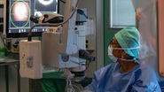
Very often, surgeons perform one cataract surgery after the other, sometimes even using several operating theaters for efficiency. The working posture in the OR can put strain especially on the neck and shoulders and result in musculoskeletal pain. Ergonomic accessories help to reduce this strain and enable surgeons to work more comfortably.
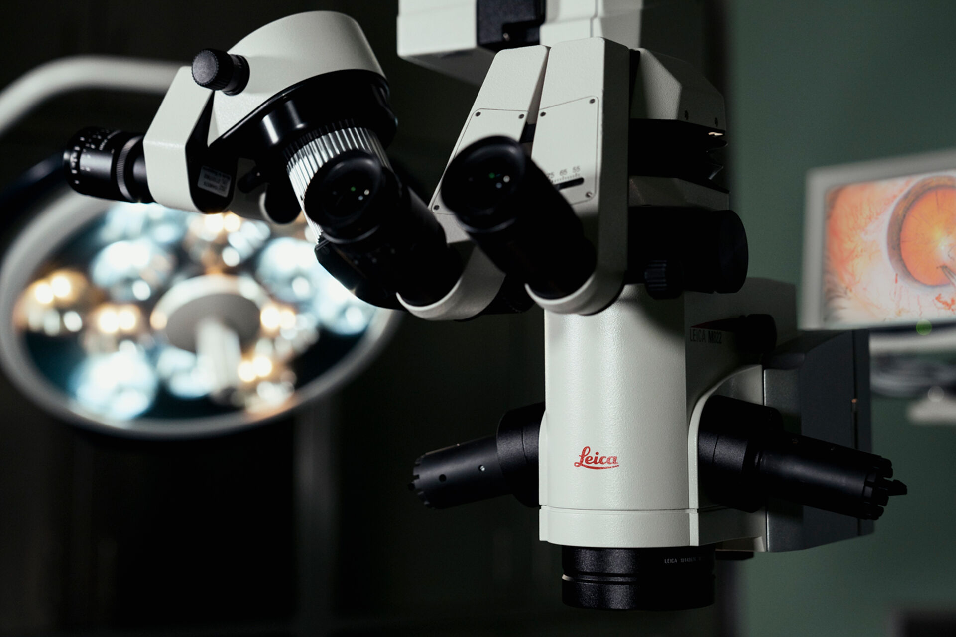
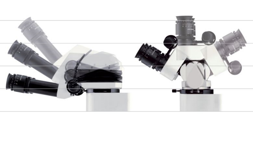
Leica surgical microscopes can be used with a variety of ergonomic binoculars which influence the viewing position during procedures. When choosing a new ophthalmic microscope, surgeons should try it out and pay special attention to the posture they work in – they will feel the benefit later.
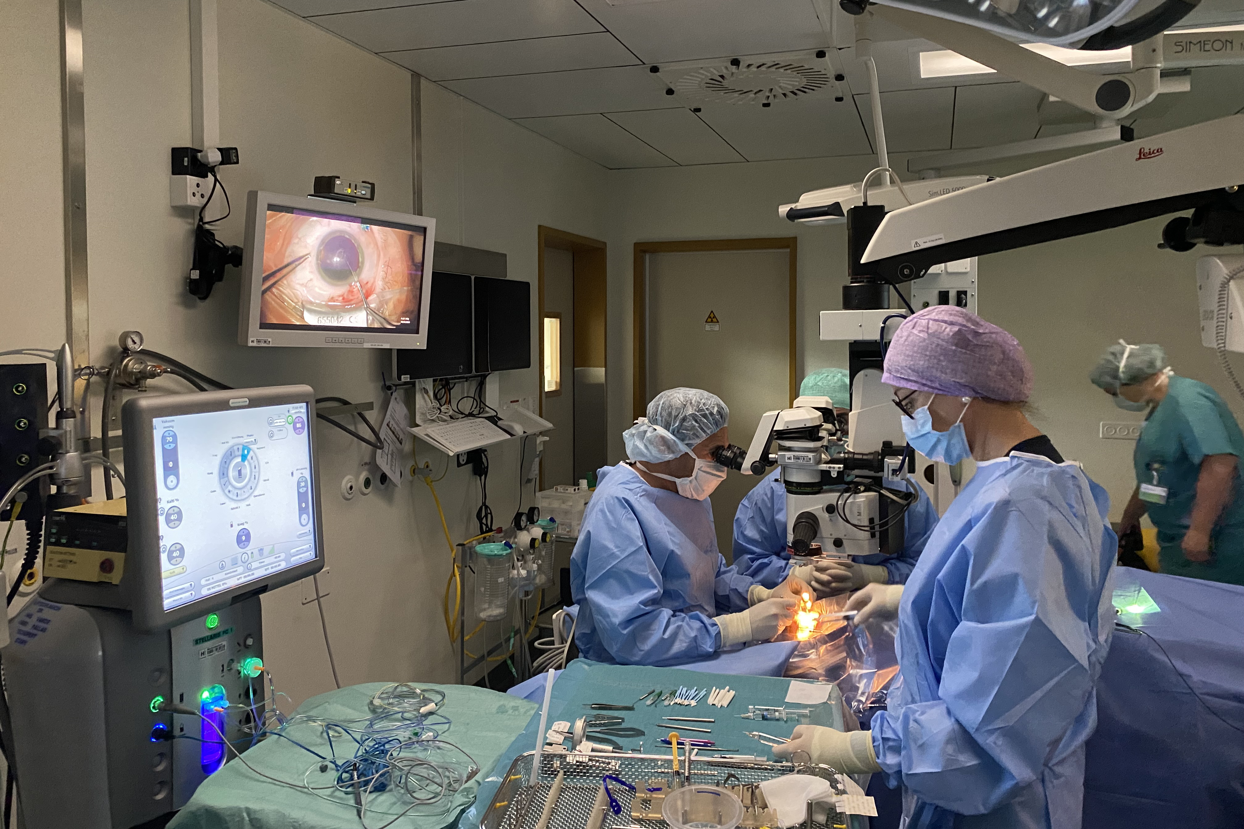
Nowadays, ophthalmic microscopes can be operated in a variety of ways to adjust to surgeon and OR team preferences. A wireless footswitch is a useful accessory. It can be programmed to save settings from several users and be operated by tapping it with a foot. This avoids unnecessary movement in the OR and reduces workflow interruptions which might be caused by setting changes.
Some microscopes offer the option to program settings such as magnification, focus, and illumination for frequently reoccurring surgical phases. Leica Microsystems offers this option in the M822 with the Step Cycle function and in the Proveo 8 with Combination Mode. This supports a smooth workflow in the OR, can reduce procedure duration and can thus enhance patient comfort. This option also enhances efficiency for the entire OR team, as the settings can be selected with the surgeon’s profile before each procedure.
Low cost of ownership, smooth maintenance workflow
Considering the budget for a new surgical microscope should also take maintenance into account. Depending on how much an ophthalmic microscope with fiber optics is used, the optical fibers may need to be exchanged. They wear with every movement of the microscope arm, as they span the distance from the microscope body to the optics carrier. This movement may result in tiny damages to the fibers, thus affecting illumination intensity over time. As a result, surgeons may need to increase the illumination values to be able to maintain the brightness and visualization quality. If the visualization quality is no longer sufficient, optical fibers may need to be exchanged.
Direct illumination reduces cost of ownership as the illumination source is in the optics carrier with no fibers needed. With a lifetime of approx. up to 50,000 hours in LED, there is no need to exchange the lamps of Leica ophthalmic microscopes during their lifetime.
If a surgical microscope is easy to clean, it takes less time to do so for the OR staff. Watch out for nooks and crannies – flat surfaces and internally routed cables are better suited to easy cleaning. Antimicrobial coating of surfaces also helps enhance hygiene. Last, but not least, anything that can be removed to be sterilized, such as handles or covers, should be made from durable materials to ensure longevity.
Upgrade options
Depending on the present and future set-up of the clinic, it may be worthwhile to consider the upgradeability of the ophthalmic microscope. E.g., if the use of the microscope shall be extended beyond cataract surgery to cover further anterior or posterior procedures, options to integrate intraoperative optical coherence tomography (OCT) may become important.
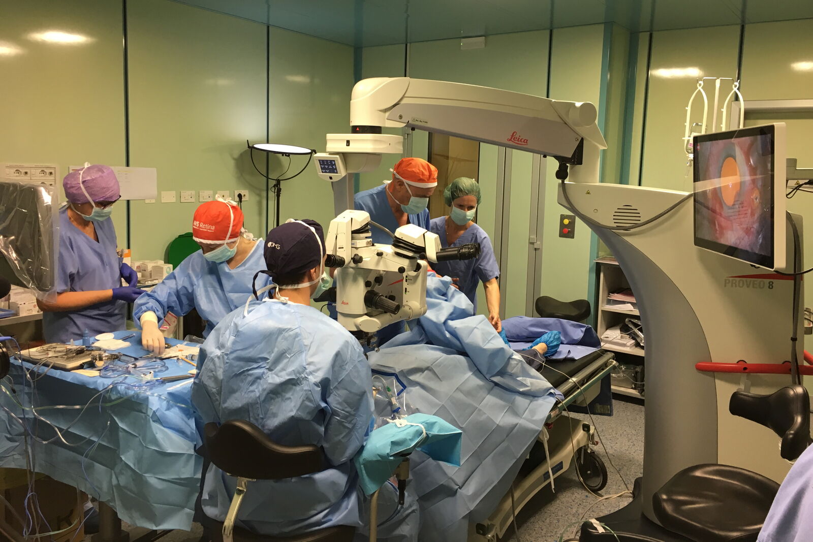
If the clinic seeks to move into teaching, integration of more extensive documentation or also teaching accessories such as viewing options for students can become relevant. Specifically for this purpose, 3D visualization technology becomes more and more frequent. Operating “heads-up” does away with the need for binoculars and enables teaching of nurses via screen. It’s worth checking whether an ophthalmic microscope can be used with a 3D visualization system. The M822 and the Proveo 8 ophthalmic microscopes by Leica Microsystems, for example, can be used with the NGENUITY® 3D Visualization System from Alcon, so that teaching takes on a new dimension.
![[Translate to chinese:] How the M822 microscope enhances surgical precision in eye surgeries - Insights from Dr. Dhami. Image courtesy of Dr. Abhinav Dhami.](/fileadmin/_processed_/d/3/csm_Dr_Abhinav_Dhami_with_M822_eb0e58ef44.jpg "[Translate to chinese:] How the M822 microscope enhances surgical precision in eye surgeries - Insights from Dr. Dhami. Image courtesy of Dr. Abhinav Dhami.")