Melinda Angus-Hill

Melinda Angus-Hill

Melinda Angus-Hill completed a Ph.D. in Oncological sciences and a postdoctoral fellowship in translational cancer research and genetics, where she developed mouse models of human disease. She was a Validation Scientist for the Cell DIVE, super-resolution and high content imaging solutions, where she represented the voice of customer perspective, and provided technical expertise and application validation support. Melinda has also led a research laboratory as a Principal Investigator and Assistant Professor at the Huntsman Cancer Institute in Utah, US, where she studied the effects of mind body medicine and stress reduction on the tumor microenvironment and tumor progression in colorectal cancer models.  
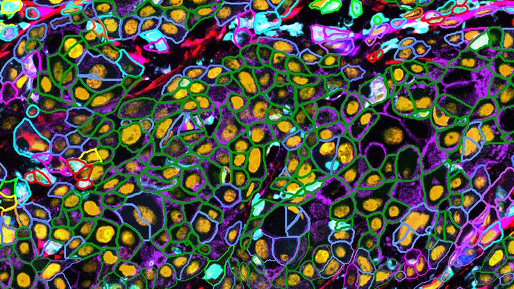
Cell counts for each biomarker were divided by total number of cells to give a percentage of biomarker positive cells out of total cells for each biomarker.

改善空间生物学研究可重复性的方法

利用自动化、高质量抗体以及经验证的多重成像工作流程,Cell DIVE能够提供可重复的实验结果。
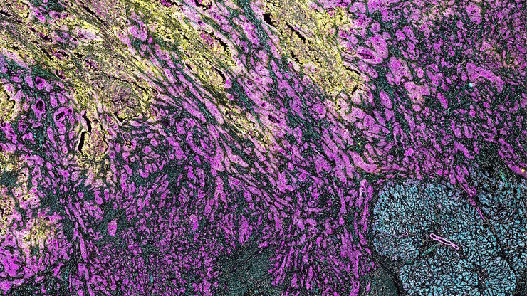
PDAC Multiplexed imaging of CST panels enables an examination of immune cell components in pancreatic ductal adenocarcinoma (IPDAC) tissue on a single slide.

表征肿瘤环境以揭示洞察和空间分辨率

肿瘤环境的表征可以为癌症进展和潜在治疗靶点提供更深入的见解。我们已经使用来自Cell Signaling Technology(CST)的各种IHC验证抗体,在胰腺癌的Cell DIVE研究中验证了30多种偶联抗体。
Pancreatic Ductal Adenocarcinoma with 5 biomarkers shown – SMA, PanCK PCK26, PanCK AE1, Vimentin, and Glut1.

借助多重成像深入了解胰腺癌的复杂性

胰腺癌是一种很难治疗的肿瘤疾病。Cell DIVE多重成像可以视觉呈现30种生物标志物以探测胰管癌的微环境。此面板可以检查肿瘤组织多个层级的问题,包括淋巴细胞、血管新生、转移、侵袭、炎症、缺氧、代谢和免疫。多重成像和分析可以对肿瘤组织中的许多生物活动提供更为深入的洞察信息,从而可以深入研究这些信息。

利用多重中频成像设计您的研究课题

多重组织分析是一种功能强大的技术,可对单个固定组织样本中的细胞类型位置和细胞类型相互作用进行比较。在多重分析研究开始之前,研究人员通常会提出以下问题: "我如何知道组织中哪些生物标记物是相关的?另外,随着研究问题的发展,我如何转向其他生物标记物?巧妙的研究设计有助于回答现有的问题,并能继续探索研究开始时并不明显的新联系。

Cell DIVE已验证的抗体将使您对实验结果产生信心

Cell DIVE超多标组织成像分析整体解决方案包括经严格验证的350+抗体资源库,高灵敏度高特异性的应用于Cell DIVE循环染色成像中。抗体验证的方法可以帮助您找到合适的抗体以及最佳的实验条件,快速的让您开展超多标成像分析的实验。抗体库中的每种抗体都经过严格的三步验证过程,(a)评估在FFPE上的表现性能;(b)确定其最佳的染色条件以及是否可用作直标抗体;(c)探究由于Cell…
Scroll to top