需要帮助?
联系当地专家,获取专业建议,帮助您选择最符合您需求和预算的解决方案。

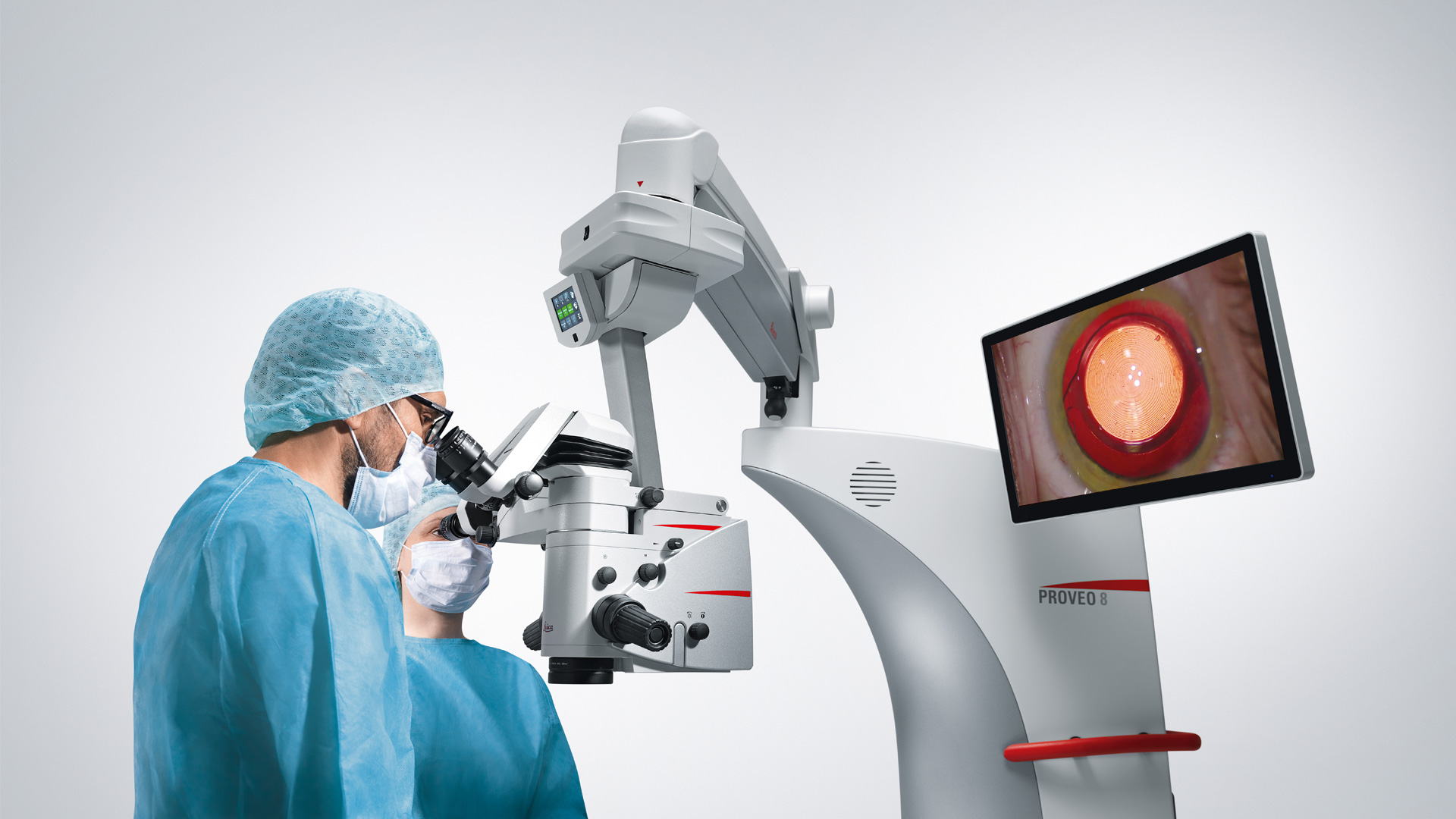
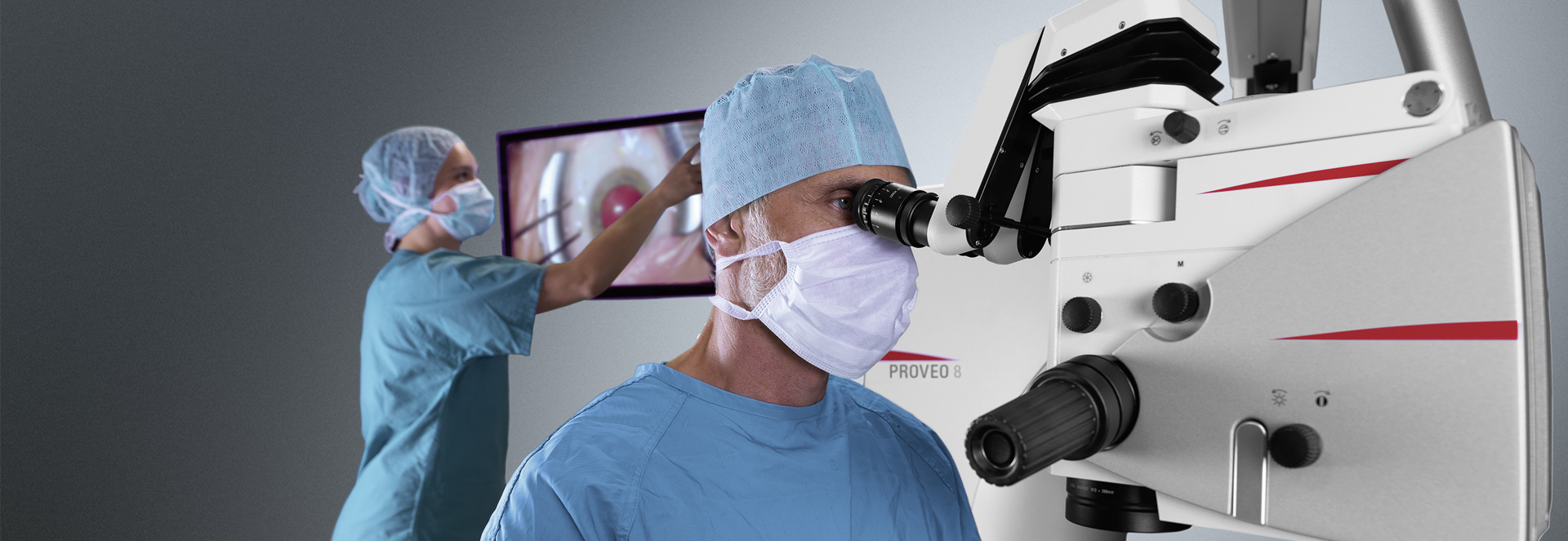
最佳照明
在撕囊和超声乳化手术中,红反射照明也能显示最精细的细节。在较低的照明水平下,红反射也能保持稳定和明亮。

不间断的手术流程
高清晰度的复消色差光学系统为眼科医生提供了大景深,因此他们无需重新聚焦即可清楚地观察。

灵活的人体工程学
可以定制眼科显微镜,以适应每个外科医生和诊所的设置,以实现无疲劳工作。
Related Articles

4 Key Benefits of 3D Digital Microscopy in Ophthalmic Surgery
3D digital visualization is rapidly transforming ophthalmic surgery. Modern 3D surgical microscopes enable surgeons to perform procedures using high-resolution digital displays rather than traditional…

Expert Techniques for Superior Visualization in Cataract Surgery
Join renowned ophthalmic surgeons, Dr. Hussein Almuhtaseb and Mr. Simon Madge, as they share their clinical expertise and real-world surgical strategies during the 2025 Online Cataract Surgery…

听听Dhami 医生关于购买眼科显微镜的专业见解
在本文中,了解来自印度北部的眼科手术顾问Abhinav Dhami医生如何使用Leica Microsystems的M822眼科显微镜来提高他的手术精确度,以及哪些关键特性使他决定购买这款眼科显微镜用于他的实践。

术中 OCT 辅助下的脱位性白内障继发房角闭合病例
了解如何在术中 OCT 的辅助下通过闭角术治疗脱位白内障,从而获得长期的良好效果,避免日后晶状体脱位。

眼科: 复杂白内障手术中的可视化
白内障手术是最常见的眼科手术。为了满足白内障手术的需要,Ozana Moraru 博士使用了 Leica Microsystems 的 M844 显微镜和 EnFocus 术中光学相干断层扫描 (OCT) 以及 3D 可视化系统。在本案例研究中,她介绍了术中光学相干断层扫描如何为标准和复杂的白内障手术病例提供有用信息。

Tawfik医生分享了他对白内障手术中水平劈核术的专业见解
据估计,每年全球约有2800万例白内障手术[1]。超声乳化术是去除白内障最常用的方法,而劈核技术在确保最佳手术效果中起着至关重要的作用。

如何选择白内障手术显微镜
掌握这些关键要素能使医生在与厂商代表洽谈时准备充分。多家企业提供设备演示服务——建议正在物色合适眼科显微镜的外科医生充分利用这一选项。
克服眼科手术挑战
涉及眼前节和眼后节的眼科手术操作可能尤其具有挑战性。良好的可视化效果是确保手术精准度和操作信心的必要条件。
Martin Spitzer 教授是德国汉堡-埃彭多夫大学医学中心眼科诊所的主任。它是德国北部最大的大学眼科诊所,每年有超过 4,800 名住院病人和近 25,000 名门诊病人。斯皮策教授和他的团队使用 Leica Proveo 8 显微镜进行诊所的所有手术。他们每年进行 8000…
先进技术在白内障和屈光手术领域的应用
在本次网络研讨会中,Thompson博士和Moshirfar博士将解释徕卡显微镜在以下手术的中作用:在LIKE手术中多焦IOL和角膜嵌体(例如Kamra和Lenticular Grafts)的置中处理。

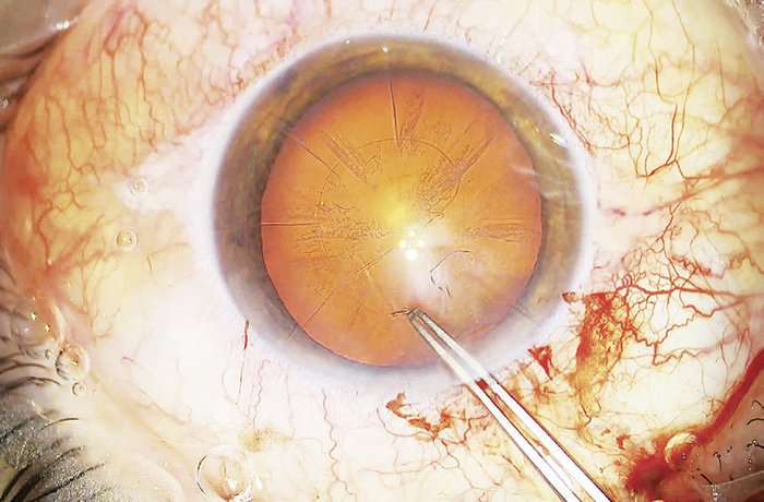
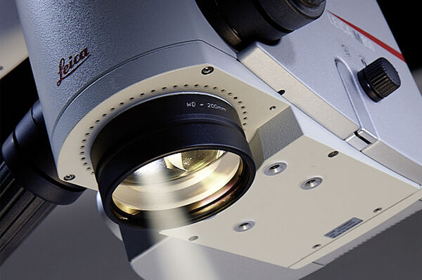
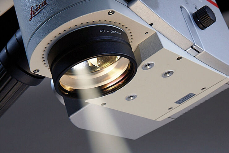
使用CoAx 4 四光束同轴立体照明技术实施的白内障手术
稳定的红光反射是白内障手术所用的眼科手术显微镜的最重要功能之一。红光反射让手术医生可以观察到晶状体结构,为其安全成功地实施手术提供清晰的视野。如何能清晰的观察到晶状体结构,特别是在手术过程中的超声乳化、晶状体摘除以及人工晶状体植入等关键阶段,始终提供稳定的红光反射,是手术显微镜面临的挑战。
但在白内障超声乳化等手术的关键阶段,传统手术显微镜的红光反射照明通常会减弱。而一种具有四条独立光路的新照明…
