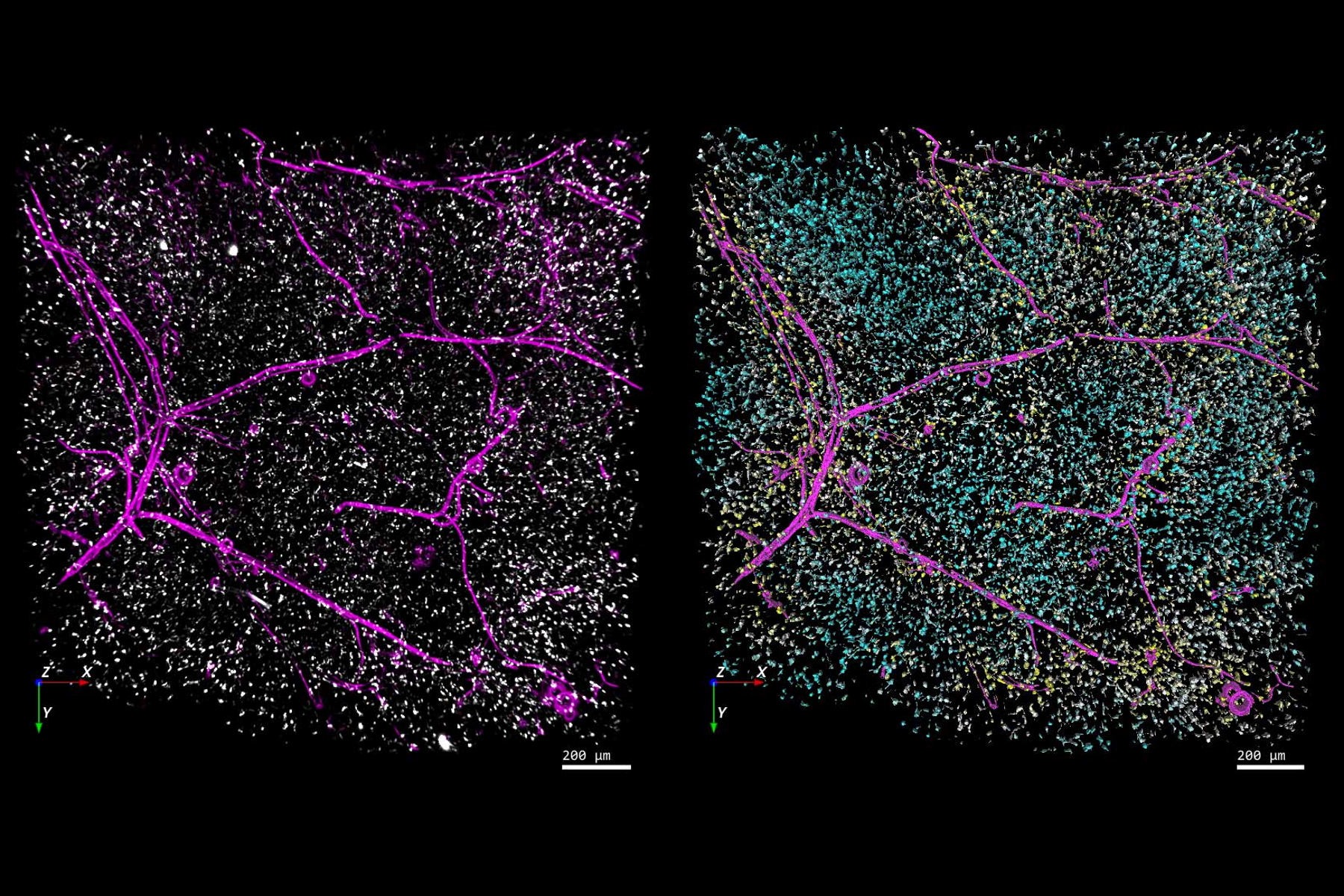
![[Translate to chinese:] Left-hand image: The distribution of immune cells (white) and blood vessels (pink) in white adipose tissue (image captured using the THUNDER Imager 3D Cell Culture). Right-hand image: The same image after automated analysis using Aivia, with each immune cell color-coded based on its distance to the nearest blood vessel. Image courtesy of Dr. Selina Keppler, Munich, Germany. Fat_tissue_THUNDER_THUNDER-Aivia_teaser.jpg](/fileadmin/_processed_/f/a/csm_Fat_tissue_THUNDER_THUNDER-Aivia_teaser_8e997b74ce.jpg "[Translate to chinese:] Blood vessels imaged with THUNDER & Aivia")

相关文章
-

A Meta-cancer Analysis of the Tumor Spatial Microenvironment
Learn how clustering analysis of Cell DIVE datasets in Aivia can be used to understand…
Apr 26, 2024Read article -
 tissue.")
Mapping the Landscape of Colorectal Adenocarcinoma with Imaging and AI
Discover deep insights in colon adenocarcinoma and other immuno-oncology realms through the potent…
Apr 26, 2024Read article -

Spatial Architecture of Tumor and Immune Cells in Tumor Tissues
Dig deep into the spatial biology of cancer progression and mouse immune-oncology in this poster,…
Apr 26, 2024Read article
相关页面
-

-

THUNDER Imaging Systems
为了解答重要的科研问题,这些系统甚至能深入原始样品中实时呈现清晰的细节,不会产生任何离焦模糊。现如今,为3D样品进行清晰成像就像使用您最喜爱的摄像头荧光显微镜一样简单。采用 Computational…
Visit related page -

想了解更多信息?
请咨询我们的专家。
您想获取专人咨询吗? Show local contacts