10-14 June 2023, Heidelberg, Germany – the course focusing on fluorescence lifetime-based readouts organized with the EMBL Imaging Centre gathered…
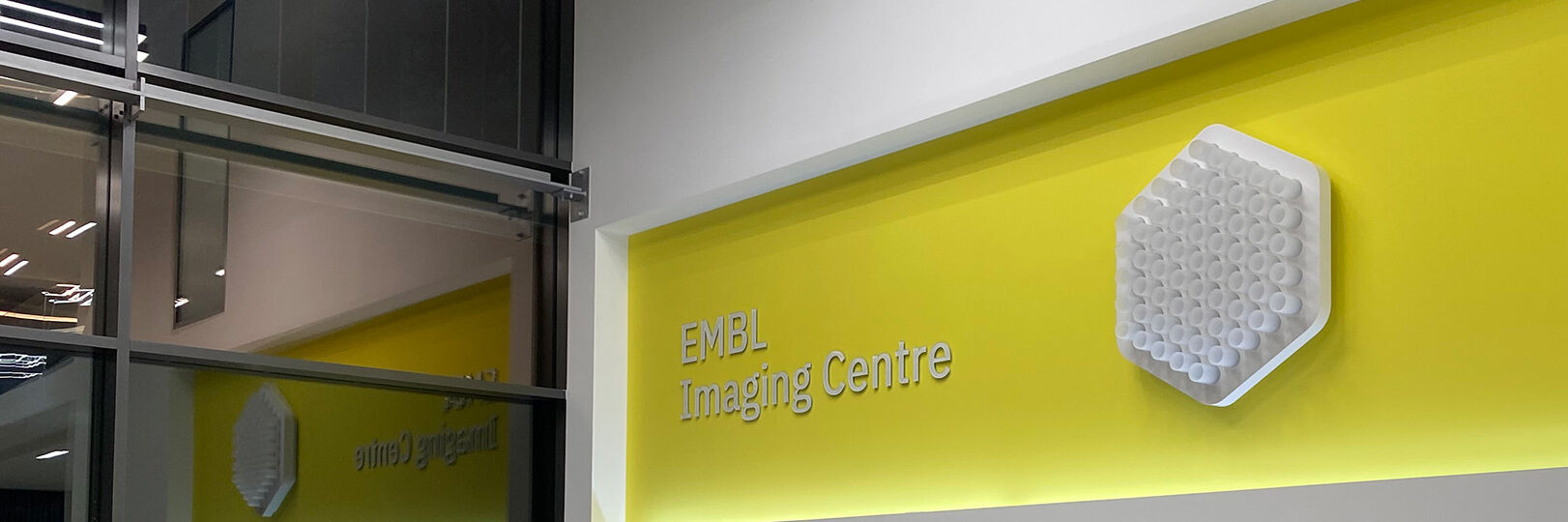
Leica and the EMBL Imaging Centre – Enabling Open Access
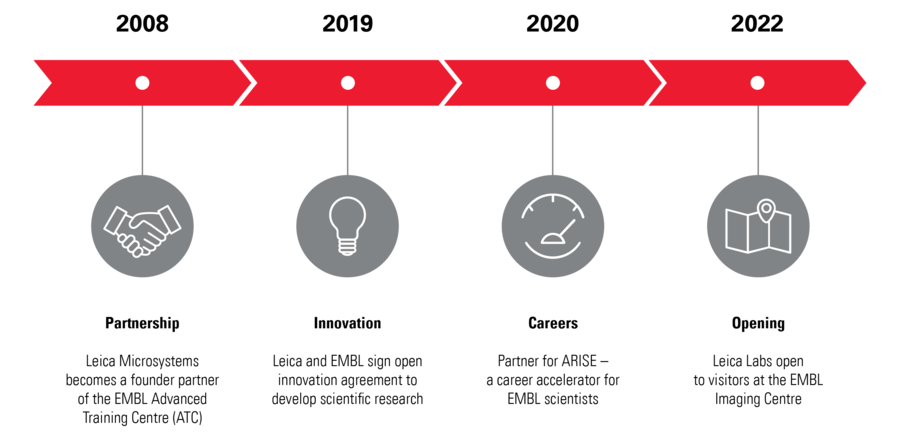
Throughout its history, Leica has enthusiastically developed relationships with academic and scientific research institutions to advance scientific understanding through microscopy. Now, thanks to our special partnership with the European Molecular Biology Laboratory (EMBL) in Heidelberg, researchers can gain access to cutting edge sample-prep and imaging technology.
By supporting EMBL’s drive to better understand the molecular basis of life through the provision of the latest technologies and expert support for a wide range of scientific and experimental services, Leica helps scientists push the boundaries of their research further and unlock greater insights.
Leica is among four industrial partners which helped to realize the vision of the EMBL Imaging Centre, which officially opened in 2022.
Find out more
Visit the EMBL IC website to see the instruments available to research applicants.
Latest Updates

The EMBO (European Molecular Biology Organization) practical course took place between the 12th and the 17th of February 2023. It covered all steps of…
 setting up the STELLARIS 8 STED for the virtual course")
Jointly organized by Leica Microsystems and EMBL, this virtual course held between the 8th and 15th of July 2022, provided attendees with deeper…
")
The EMBL Imaging Centre was inaugurated on the 30th of June 2022, welcoming representatives from politics, industry and research. The center was…

The EMBL in Heidelberg hosted its first Scientific Symposium at the EMBL Imaging Centre on the 30th of May 2022, featuring talks about leading edge…
Interviews
Interview with Yassin Harim, PhD Student at German Cancer Research Center, Heidelberg
Gain insights into 3D- whole mouse brain imaging using multicolor immunofluorescence! According to our guest user from the German Cancer Research Center, using THUNDER Imager Live cell at the EMBL IC was “…the perfect solution to get very high-quality images and also to spend little time on imaging because it's just so fast to acquire each individual slide”.
Interview with Virginia Pierini – Service Manager EMBL IC, Heidelberg. Virginia Pierini is supporting the operation of the EMBL Imaging Centre regarding all its services, with a special focus on users. She is the IC’s point of contact for all users, providing help around access procedures, the project execution as well as user training.
Interview with Virginia Pierini – Service Manager EMBL IC, Heidelberg.
Virginia Pierini is supporting the operation of the EMBL Imaging Centre regarding all its services, with a special focus on users. She is the IC’s point of contact for all users, providing help around access procedures, the project execution as well as user training.
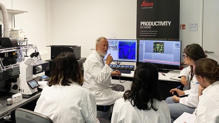
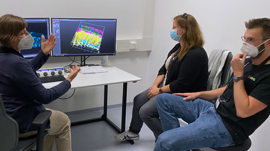
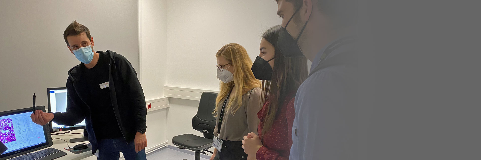
The EMBL Imaging Centre houses the latest state-of-the-art instrumentation from Leica and others, as well as those developed in EMBL research groups.
The EMBL IC offers researchers access to scientific experts from both academia as well as industry, providing its users with the opportunity to perform cutting-edge science with a suite of tools and support that are unavailable to most scientists.
Leica experts are on-site permanently at the EMBL IC to empower researchers to use the data from its advanced imaging systems in order to achieve groundbreaking insights.
Apply now
Bring your research project to EMBL and get support by the Leica Microsystems application specialists.
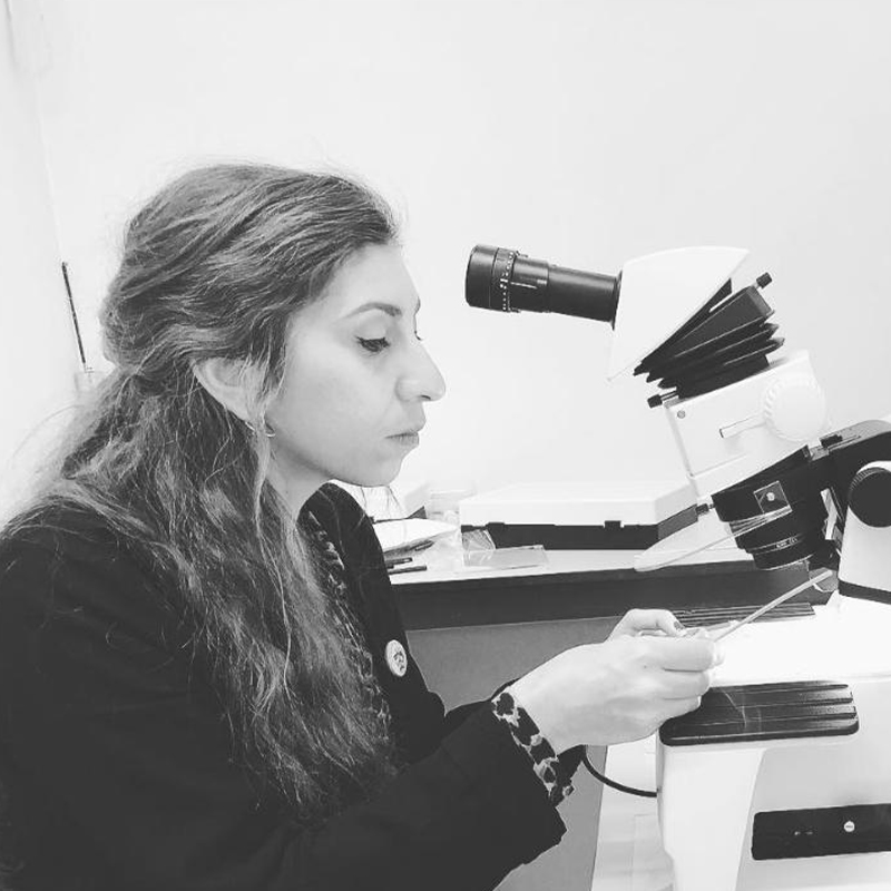
Meet the Leica Team at the EMBL IC

Robert Kirmse
Robert received his PhD from the DKFZ and University of Heidelberg. As post-doc, he worked on tumor cell invasion at BioQuant, Heidelberg and in cryo EM at the University of Colorado, Boulder. He joined Leica in 2019 as senior manager for sample preparation and site lead in Vienna. Since October 2022 he leads Leica’s EMBL IC team for Scientific Innovation.

Lynne Turnbull
Lynne received her PhD in Sydney, Australia and postdoctoral training in San Francisco and Melbourne. She used imaging to understand bacterial biofilms and how bacteria move. At UTS (Sydney), Lynne managed the Microbial Imaging Facility. Since 2021 Lynne has been a Principal Scientist with Leica Microsystems at the EMBL Imaging Center in Heidelberg.

Andrea Mülter
Andrea joined Leica as product manager. After leading the global application team, she currently manages Leica’s knowledge program and strategic relations with leading scientists. She obtained her PhD at NIH with Jennifer Lippincott-Schwartz and then worked with Ursula Klingmüller at DKFZ in Heidelberg to study signal transduction by systems biology.
Andreia Pinto
Andreia worked as an electron microscopy specialist in Lisbon for 11 years. In 2019, she moved to London to finish her PhD and work in the fields of AI and Covid-19. Currently, she is an Advanced Workflow Specialist at Leica Microsystems and is based at the EMBL Imaging Centre in Heidelberg.

Martin Fritsch
Martin is a broadly trained biologist with a PhD in zoology. During his post-doc in evolutionary and developmental biology (EvoDevo) he focused on non-model organism invertebrates. In 2017, he joined Leica as a sales specialist for confocal solutions and is now an advanced workflow specialist for advanced point scanning and fluorescence microscopes.
EMBL collaboration – a timeline
EMBL related articles

Benefits of TauContrast to Image Complex Samples
In this interview, Dr. Timo Zimmermann talks about his experience with the application of TauSense tools and their potential for the investigation of demanding samples such as thick samples or…

New Imaging Tools for Cryo-Light Microscopy
New cryo-light microscopy techniques like LIGHTNING and TauSense fluorescence lifetime-based tools reveal structures for cryo-electron microscopy.

How to Target Fluorescent Structures in 3D for Cryo-FIB Milling
This article describes the major steps of the cryo-electron tomography workflow including super-resolution cryo-confocal microscopy. We describe how subcellular structures can be precisely located in…

Precise 3D Targeting for EM Imaging - Access What Matters
Find out how the seamless cryo-electron tomography workflow Coral Cryo uses confocal super resolution to target your structure of interest more precisely.
 embryo, from sphere stage to somite stages.")
Studying Early Phase Development of Zebrafish Embryos
VIDEO ON DEMAND - This second edition of MicaCam focuses on combining widefield and confocal imaging to study the early-stage development of zebrafish embryos (Danio rerio), from oocyte to…

How Marine Microorganism Analysis can be Improved with High-pressure Freezing
In this application example we showcase the use of EM-Sample preparation with high pressure freezing, freeze substiturion and ultramicrotomy for marine biology focusing on ultrastructural analysis of…