
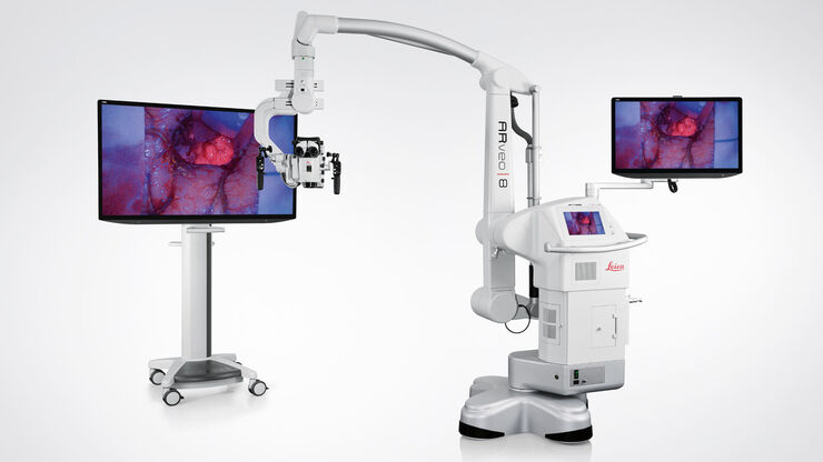
Evolved ARveo 8 Digital Visualization Microscope for Neurosurgery
Not all products or services are approved or offered in every market, and approved labelling and instructions may vary between countries. Please contact your local representative for further information. FDA 510(k) clearance for GLOW400 is pending.
Experience a new level of neurosurgical visualization with the evolved ARveo 8 digital visualization microscope. It is available with a clinical 3D application that will transform your brain tumor surgery visualization. The all-in-one surgical visualization headset, MyVeo, will take you beyond what you’ve imagined with a single, integrated view of clinical data right in front of your eyes.
ARveo 8 evolves continuously
Get ready for a new level of continuous access to digital capabilities that are pushing the boundaries of neurosurgery as you know it.
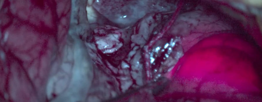
Augmenting brain tumor surgery
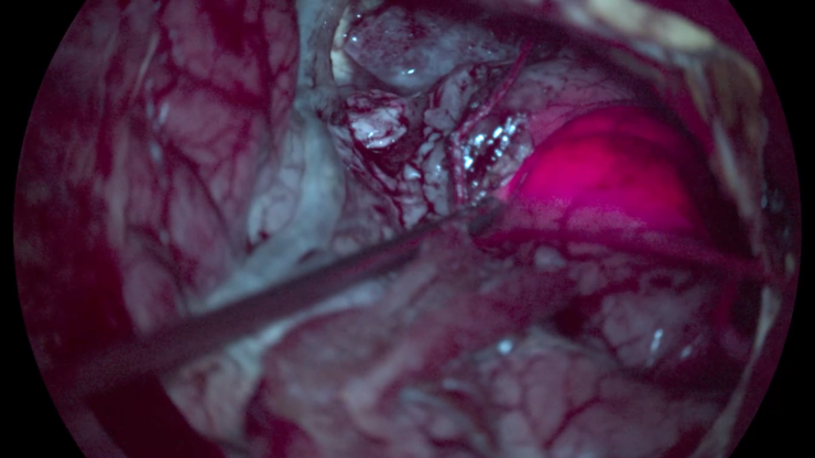
Get a real-time augmented view with 3D depth perception of suspected grade III and grade IV glioma tissues with the GLOW400 Augmented Reality fluorescence application for brain tumor surgery.
The application transforms your brain tumor visualization:
- Anatomy view: see enhanced anatomical details surrounding the fluorescent-marked tumor, even showing details such as vessels or bleeding.
- Highlighted Fluorescence view: Get a broader representation of fluorescence intensities, that you may have previously missed.

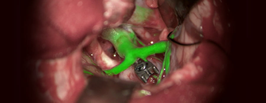
Augmenting vascular neurosurgery: anatomy and blood flow in one image
The GLOW800 AR fluorescence application for vascular neurosurgery allows you to observe cerebral anatomy and blood flow in white light.
Forget yesterday's need to recall and reconcile black and white NIR blood flow video with the natural anatomical view—enjoy one single view of anatomy and blood flow.
Stay orientated thanks to digital 3D depth perception without dark peripheries Work confidently with the GLOW800 application for removal of AVMs, clipping of aneurysms, performance of a bypass or a microvascular decompression


Look beyond what you’ve imagined
The MyVeo all-in-one surgical visualization headset for the ARveo 8 digital visualization microscope frees you from the microscope by unifying essential clinical data right in front of your eyes, helping you and your team to stay focused and increase physical comfort.
Three MyVeo users can simultaneously experience real-time surgery, which is especially useful and powerful for teaching and learning.

Experience enhanced efficiency across different procedures
The ARveo 8 surgical microscope is amazingly versatile. This is based on the microscope’s great range of movement, large working distance, tilting range of the optics carrier, and an extensive overhead reach.
- Achieve a comfortable upright working posture for both the main and opposite assistant thanks to the design of the optics carrier
- Benefit from up to 600 mm working distance, providing more space to pass and maneuver instruments, e.g., in spine surgery

Cleverly enhanced optical visualization
The ARveo 8 optics have groundbreaking innovation at its heart: FusionOptics. Uniting an enhanced depth of field with high resolution, FusionOptics technology delivers enhanced visualization. When combined with 400 W xenon light and Small Angle Illumination, the result is a deep, bright view, in full focus through the oculars.
FusionOptics Technology
- Two separate beam paths
- One beam path provides high resolution
- The other beam path provides depth of field
- The brain merges the two images into a single optimal spatial image

Single graphical user interface for microscope operation and image acquisition
The ARveo 8 GUI is designed to be self-explanatory for all members of the OR team. It guides you through setup of the microscope, allows for intraoperative adjustments on the fly, enables image acquisition and transfer. And finally, it serves as an additional monitor to show the microscope image.
Easy operation
- Select and define different user roles and rights
- Password protect default configurations and individual user settings, e.g., GLOW800 visualization
- Increase cybersecurity with secure patient and user data
Easy imaging
- Record video and images in 2D or 3D quality utilizing a high-compression 2 TB storage space
- Quickly store images and export via USB and Ethernet to your hospital network
- Optimized data processing and connectivity for PACS and DICOM

Adapt to new technologies at your own pace
Freely select from three interchangeable viewing options: choose traditional oculars*, 3D heads-up monitors, or the most advanced MyVeo headset. You also have the flexibility to use each viewing option interchangeably.
Access the latest technology without replacing your neurosurgical visualization microscope. The EnhancePath concept, an essential part of the ARveo 8 ecosystem, allows you to seamlessly evolve into the future of digital surgery.
*Ocular view does not show GLOW AR view

Easily connect to compatible surgical devices
The ability to combine preoperative images with intraoperative imaging can be essential during procedures. You can use image-guided surgery (IGS) systems to augment your microscope view by adding anatomical and functional data onto your microscope’s white light and the fluorescence view. The evolved ARveo 8 is compatible with neuro-navigation systems of leading manufacturers.
ARveo 8 also provides technical compatibility with KARL STORZ® video systems.

Navigation-controlled robotics
The ARveo 8 digital visualization microscope for neurosurgery enables robotic alignment of the microscope’s optics carrier via the Brainlab IGS system.
- Keep your image in focus during the entire neurosurgery, thanks to the tip focus function of the latest Cranial Navigation Software from BrainLab.
- Rest assured that you always have a centered view despite microscope movement thanks to “follow tip” or “move to pin” functions.
Fluorescence options
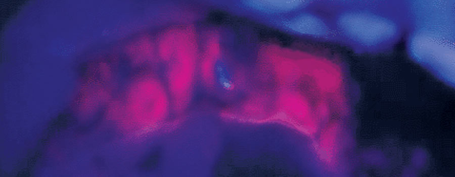
FL400 oncological fluorescence
The fluorescence module FL400 is used during open neurosurgery in conjunction with the active substance 5 aminolevulinic acid (5-ALA). It supports resection by allowing differentiation of tumor tissue from healthy brain tissue.
* Please contact Leica Microsystems Regulatory Affairs for cleared indications and registration status of the product in your region
FL560 fluorescence
FL560 allows observation of fluorophores with an excitation range between ~460 nm and ~500 nm. It allows you to view non-fluorescent tissue in natural color and simultaneously observe fluorescence in a bright yellowish-green color.
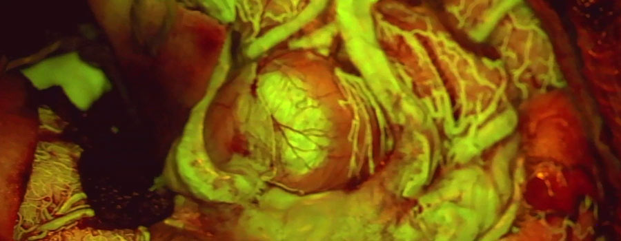
GLOW800 AR fluorescence application for vascular surgery
GLOW800 takes the high contrast of NIR imaging with ICG and combines it with white light. The result is a single view of natural-colored anatomy, augmented by real-time vascular flow visualized digitally with 3D depth perception.
GLOW400 AR fluorescence application for brain tumor surgery
Make more confident surgical decisions during suspected grade III and IV glioma surgeries with GLOW400, by seeing clearer anatomical structures surrounding the fluorescent marked tumor. GLOW400 also provides a view that reveals lower-intensity fluorescence signals that you may have previously missed.

Interested to know more?
Talk to our experts.
Do you prefer personal consulting? Show local contacts