联系我们
联系当地专家,获取有关符合您需求和预算的专家建议
相关文章

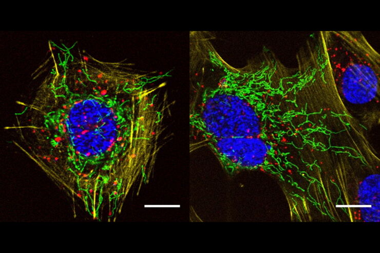
Extended Live-cell Imaging at Nanoscale Resolution
Extended live-cell imaging with TauSTED Xtend. Combined spatial and lifetime information allow super-resolution microscopy at extremely low light dose.
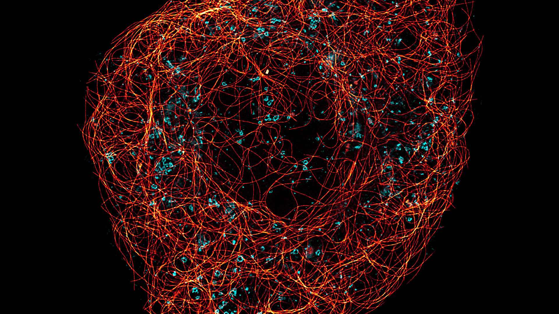
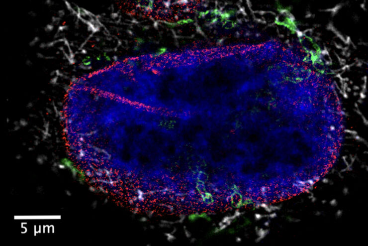
, actin network (ATTO 647N), and nuclear pore basket (CF 680R).")
The Guide to STED Sample Preparation
This guide is intended to help users optimize sample preparation for stimulated emission depletion (STED) nanoscopy, specifically when using the STED microscope from Leica Microsystems. It gives an…
![[Translate to chinese:] Five-color FLIM-STED](/fileadmin/_processed_/a/3/csm_5-color_FLIM-STED_9d88340e60.jpg "[Translate to chinese:] Five-color FLIM-STED")
采用单损耗激光的五色FLIM-STED显微镜
网络研讨会,内容涉及使用单一损耗激光和荧光寿命phasor分离技术的五色STED技术。
A Versatile Palette of Fluorescent Probes
Researchers at the Max Planck Institute for Medical Research in Heidelberg have developed a general strategy to synthesize live-cell compatible fluorogenic probes, and the result are the new MaP (Max…
Benefits of Combining STED and Lifetime
In this interview, Professor Alberto Diaspro talks about the advantages of the White Light Laser and the TauSTED capabilities of STELLARIS 8 STED. He speaks about his experience with the confocal…
透明介质对组织透明度和收缩的影响
本研究通过比较新鲜解剖的双翅目昆虫脑与其透明化处理后的等效物,全面评估了不同透明化介质对组织透明度和收缩率的影响。组织透明化处理结合光片显微技术,已成为全器官三维成像和定量的强大工具。由于组织透明化处理有助于在亚细胞水平上对完整组织进行光学成像,它有潜力揭示如大脑等复杂器官以前未见的细节。为了对厚样本进行高分辨率成像,透明化介质需要与高数值孔径(NA)物镜使用的浸没液具有匹配的高折射率。这一点对于…
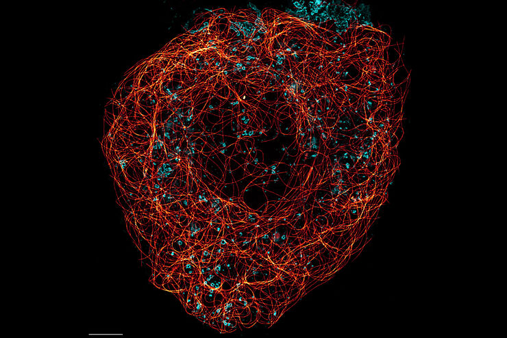
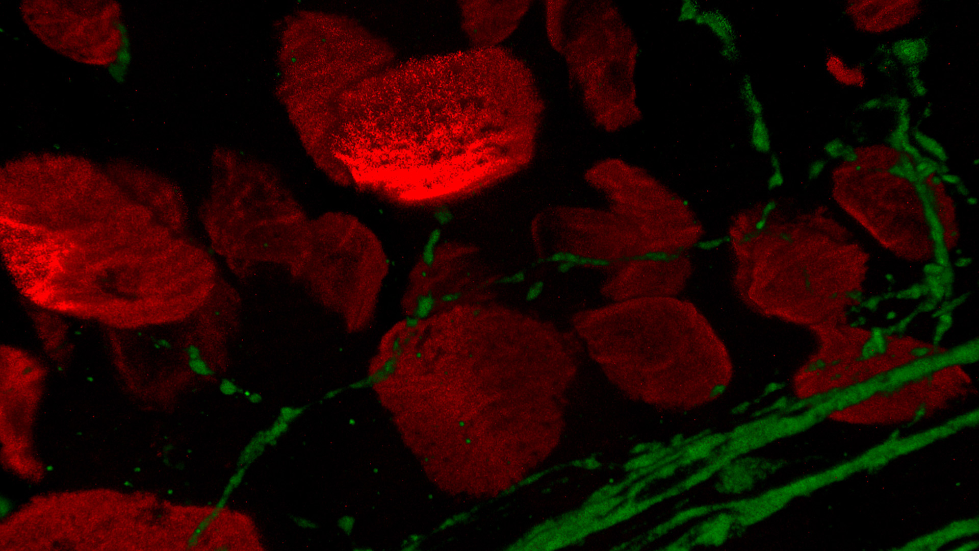
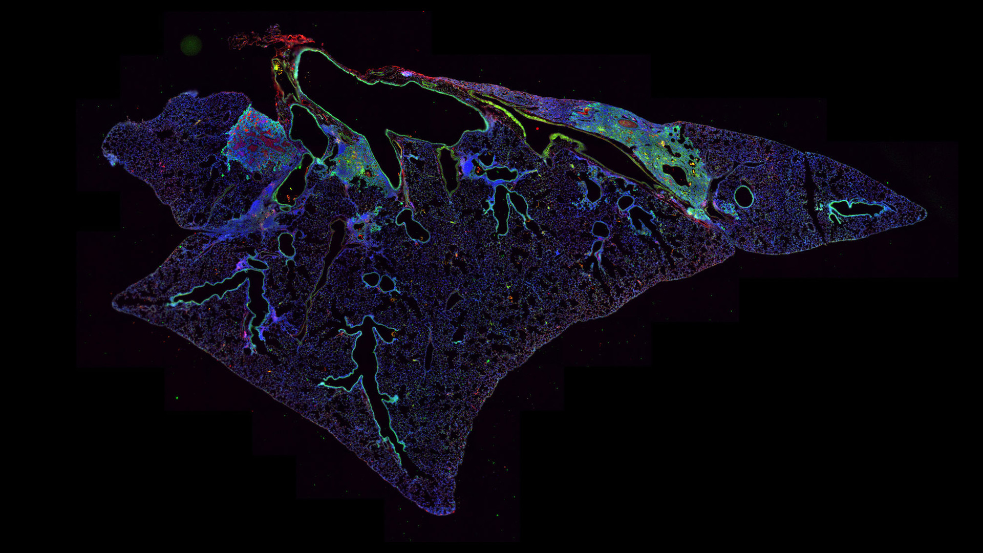
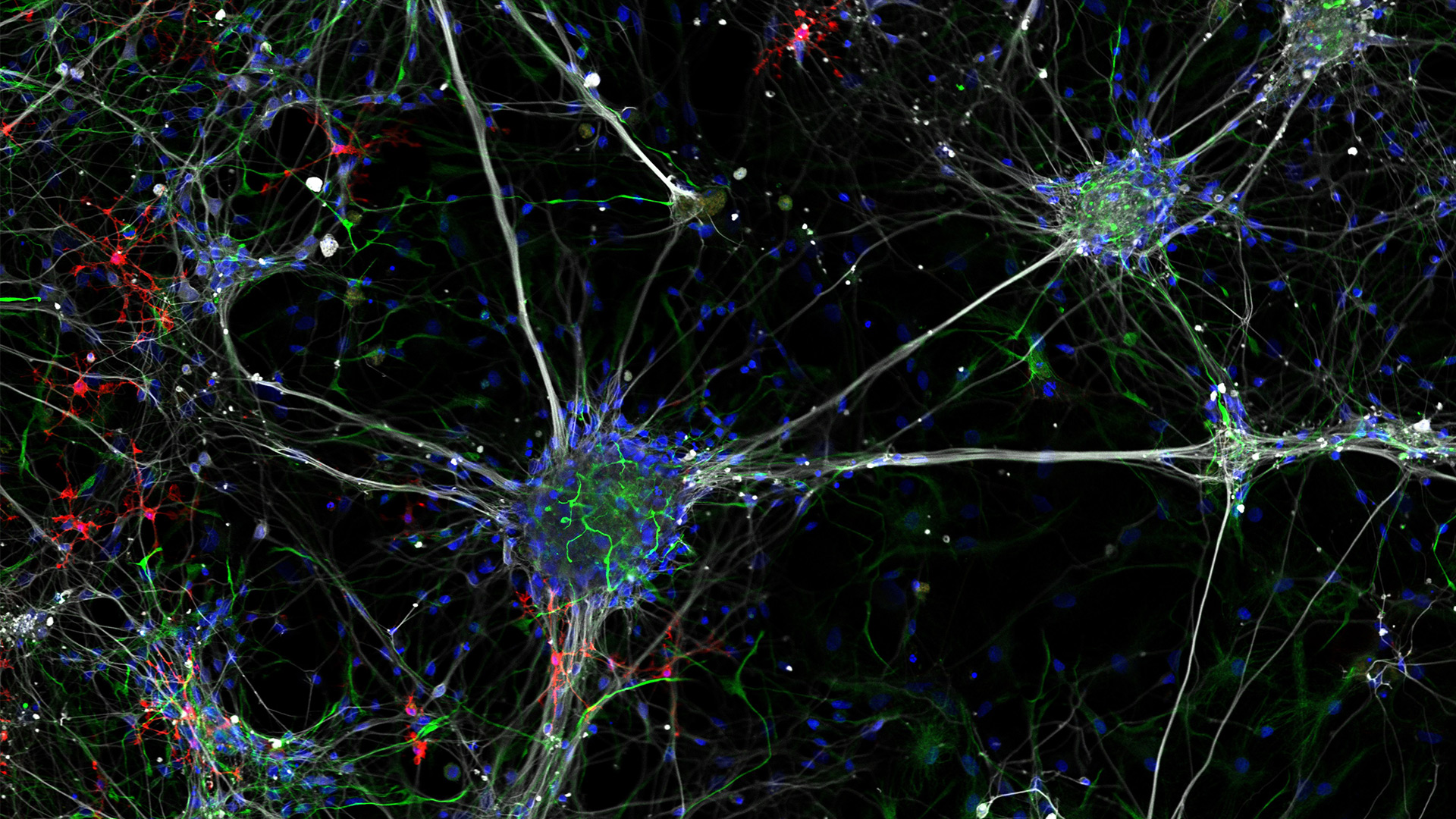
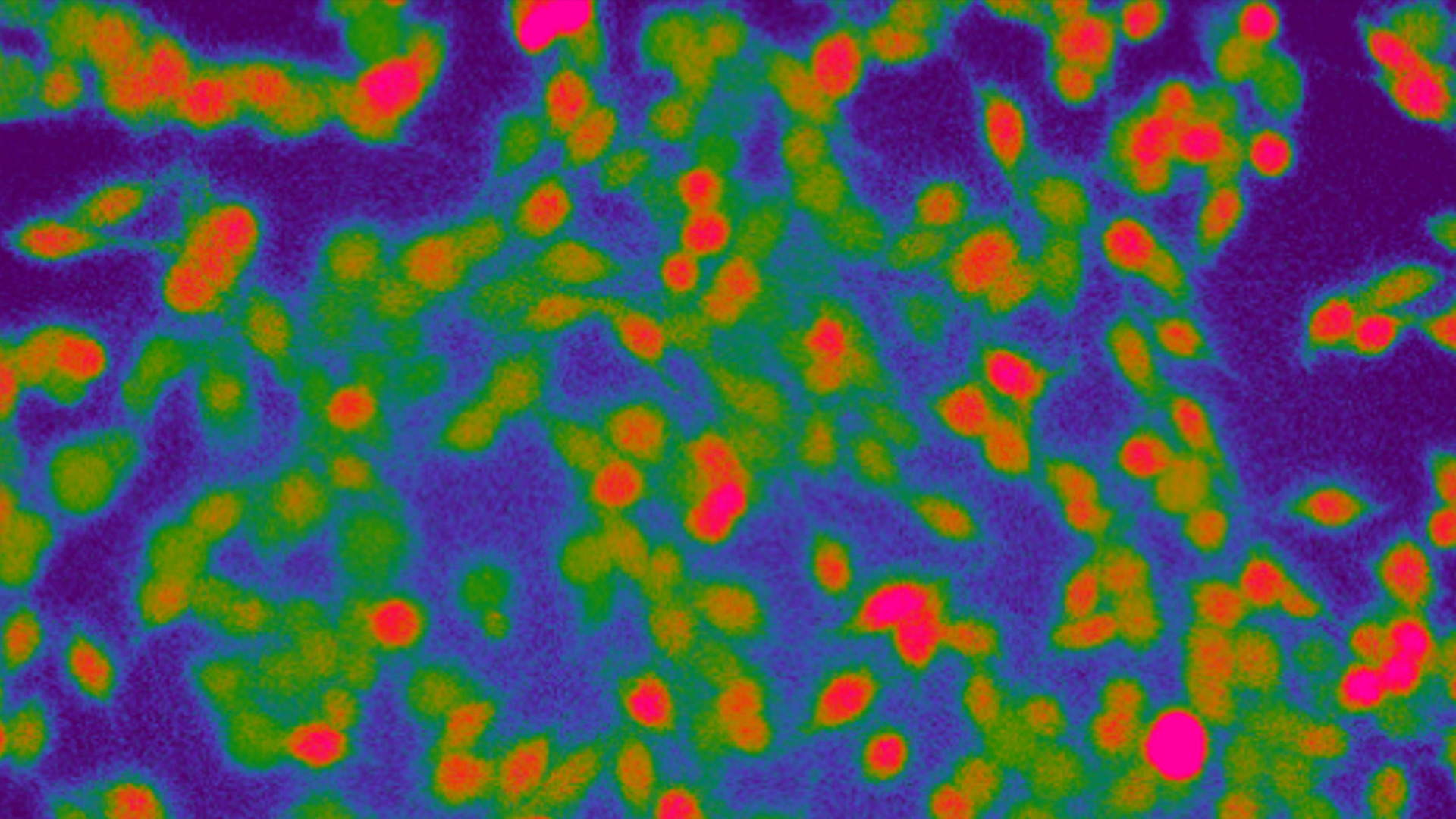
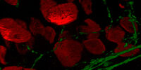
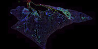
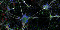
超分辨率显微镜图片库
由于光的衍射极限,传统共聚焦显微镜无法分辨约240纳米以下的结构。当需要提高分辨率以研究衍射极限尺度以下的结构和分子事件时,会使用超分辨率显微镜技术,如STED、PALM或STORM,或某些解卷积处理方法。
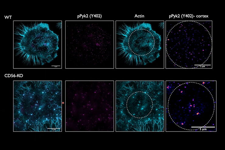
Regulators of Actin Cytoskeletal Regulation and Cell Migration in Human NK Cells
Dr. Mace will describe new advances in our understanding of the regulation of human NK cell actin cytoskeletal remodeling in cell migration and immune synapse formation derived from confocal and…

显微镜在病毒学中的应用
引起新型冠状病毒肺炎(Covid-19)的冠状病毒SARS-CoV-2肆虐全球并影响了我们生活的方方面面。对于免疫和治疗方法的搜索研究(即如何抗击该病毒)成为了2020年全人类的第一要务。显微镜在这类研究中起着重要作用。为了了解受体结合、基因组释放、复制、装配和病毒出芽的基本原理以及我们的免疫系统效应,可以使用不同的方法和显微镜。本文概述了为什么显微镜是病毒学和感染生物学的重要工具,并举例说明了不…
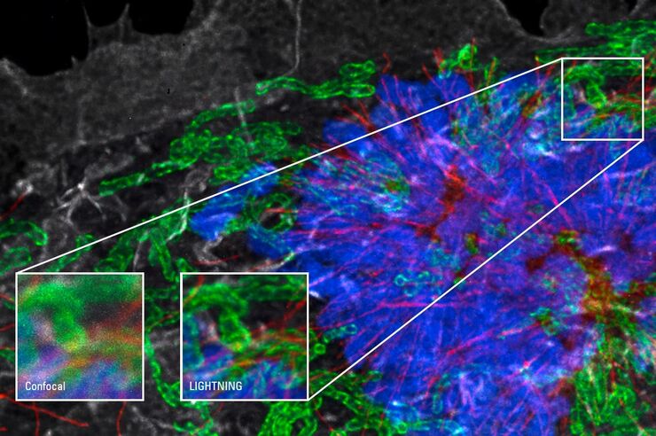
See More Than Just Your Image
Despite the emergence of new imaging methods in recent years, true 3D resolution is still achieved by Confocal Laser Scanning Microscopy (CLSM). Through a combination of novel, extremely fast scanning…

想了解更多信息?
请咨询我们的专家。
您想获取专人咨询吗? Show local contacts