Loading...

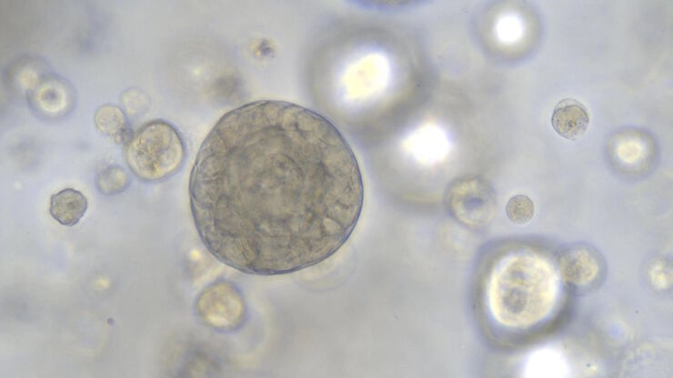
Overcoming Observational Challenges in Organoid 3D Cell Culture
Learn how to overcome challenges in observing organoid growth. Read this article and discover new solutions for real-time monitoring which do not disturb the 3D structure of the organoids over time.
Loading...
![[Translate to chinese:] Image of burrs (red arrows) at the edge of a battery electrode acquired with a DVM6 digital microscope. [Translate to chinese:] Image of burrs (red arrows) at the edge of a battery electrode acquired with a DVM6 digital microscope.](/fileadmin/_processed_/7/6/csm_Burrs_at_the_edge_of_a_battery_electrode_effd6b277b.jpg)
电池制造过程中的毛刺检测
毛刺是电池电极片边缘可能出现的缺陷,例如在制造过程中的分切环节。它们可能会因诸如短路等故障导致电池性能下降,并引发安全和可靠性问题。毛刺检测是电池生产质量控制的重要部分,对于生产具有可靠性能和寿命的电池至关重要。通过适当照明的光学显微镜可以在生产过程的关键步骤中快速可靠地对电极上的毛刺进行视觉检测。
Loading...

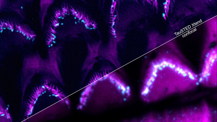
Extended Live-cell Imaging at Nanoscale Resolution
Extended live-cell imaging with TauSTED Xtend. Combined spatial and lifetime information allow super-resolution microscopy at extremely low light dose.
Loading...

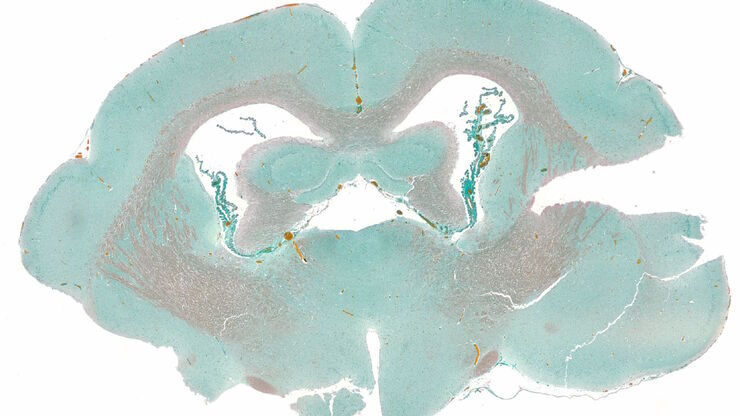
How to Streamline Your Histology Workflows
Streamline your histology workflows. The unique Fluosync detection method embedded into Mica enables high-res RGB color imaging in one shot.
Loading...
![[Translate to chinese:] Image of an integrated-circuit (IC) chip cross section acquired at higher magnification showing a region of interest. [Translate to chinese:] Image of an integrated-circuit (IC) chip cross section acquired at higher magnification showing a region of interest.](/fileadmin/_processed_/a/c/csm_IC_chip_cross_section_20a5502a5d.jpg)
横截面切片法分析IC芯片的结构与化学成分
从本文中了解如何通过横截面分析法对集成电路 (IC) 芯片等电子元件进行有效的结构和元素分析。探索如何通过研磨系统进行铣削、锯切、磨削和抛光工艺以及用于同时进行目视检测和化学分析的二合一解决方案来完成的。可针对电子行业的各种工作流程和应用实现快速、详细的材料分析,包括竞争分析、质量控制 (QC)、故障分析 (FA) 以及研发 (R&D)。
Loading...

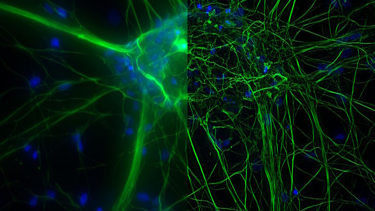
What are the Challenges in Neuroscience Microscopy?
eBook outlining the visualization of the nervous system using different types of microscopy techniques and methods to address questions in neuroscience.

![[Translate to chinese:] THUNDER image of brain-capillary endothelial-like cells derived from human iPSCs (induced pluripotent stem cells) where cyan indicates nuclei and magenta tight junctions. [Translate to chinese:] THUNDER image of brain-capillary endothelial-like cells derived from human iPSCs (induced pluripotent stem cells) where cyan indicates nuclei and magenta tight junctions.](/fileadmin/_processed_/8/e/csm_Brain-capillary_endothelial-like_cells_derived_from_human_iPSCs_b5dcf04bc9.jpg)
![[Translate to chinese:] Murine esophageal organoids (DAPI, Integrin26-AF 488, SOX2-AF568) imaged with the THUNDER Imager 3D Cell Culture. Courtesy of Dr. F.T. Arroso Martins, Tamere University, Finland. [Translate to chinese:] Murine esophageal organoids (DAPI, Integrin26-AF 488, SOX2-AF568) imaged with the THUNDER Imager 3D Cell Culture. Courtesy of Dr. F.T. Arroso Martins, Tamere University, Finland.](/fileadmin/_processed_/f/f/csm_THUNDER_Imager_3D_Cell_Culture_Murine-esophageal-organoid_LVCC_9bc587500f.jpg)
![[Translate to chinese:] Water Flea Daphnia imaged by Electron Microscopy. Courtesy of Mag. Dr. Gruber, University of Vienna, Austria. [Translate to chinese:] Water Flea Daphnia imaged by Electron Microscopy. Courtesy of Mag. Dr. Gruber, University of Vienna, Austria](/fileadmin/_processed_/e/d/csm_EM_Application_Water_Flea_Daphnia_7abc89f4fb.jpg)