
![[Translate to chinese:] Murine esophageal organoids (DAPI, Integrin26-AF 488, SOX2-AF568) imaged with the THUNDER Imager 3D Cell Culture. Courtesy of Dr. F.T. Arroso Martins, Tamere University, Finland.](/fileadmin/_processed_/f/f/csm_THUNDER_Imager_3D_Cell_Culture_Murine-esophageal-organoid_LVCC_9bc587500f.jpg "[Translate to chinese:] Murine esophageal organoids (DAPI, Integrin26-AF 488, SOX2-AF568) imaged with the THUNDER Imager 3D Cell Culture. Courtesy of Dr. F.T. Arroso Martins, Tamere University, Finland.")
Loading...
![[Translate to chinese:] Brain organoid section (DAPI) acquired using THUNDER Imager Live Cell. Image courtesy of Janina Kaspar and Irene Santisteban, Schäfer Lab, TUM.](/fileadmin/_processed_/2/7/csm_Tilescan_of_brain_organoid_section_46d510ba4e.jpg "[Translate to chinese:] Brain organoid section (DAPI) acquired using THUNDER Imager Live Cell. Image courtesy of Janina Kaspar and Irene Santisteban, Schäfer Lab, TUM.")
研究大脑健康的成像类器官模型
小胶质细胞是特化的脑驻留免疫细胞,在大脑发育、平衡和疾病中发挥着至关重要的作用。然而,到目前为止,模拟人脑环境与小胶质细胞之间相互作用的能力还非常有限。
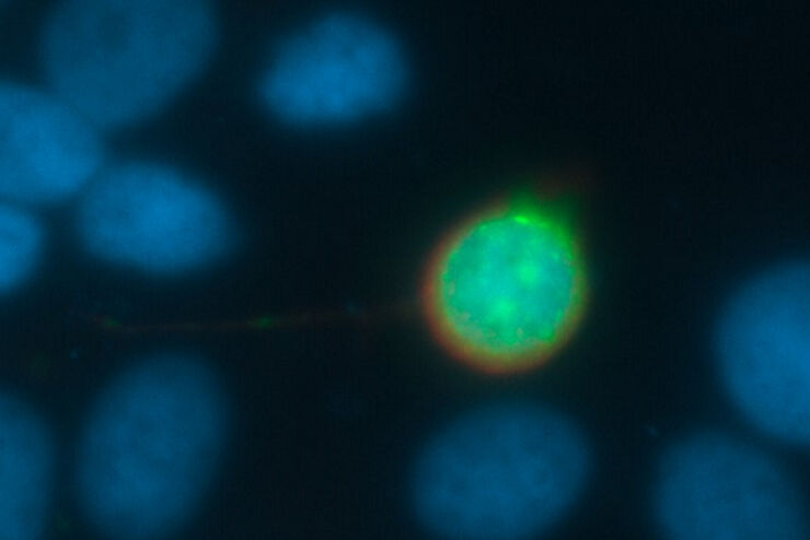
![[Translate to chinese:] Mouse cortical neurons. Transgenic GFP (green). Image courtesy of Prof. Hui Guo, School of Life Sciences, Central South University, China](/fileadmin/_processed_/2/a/csm_THUNDER_Imager_Mouse_cortical_neuron_9d4054774d.jpg "[Translate to chinese:] Mouse cortical neurons. Transgenic GFP (green). Image courtesy of Prof. Hui Guo, School of Life Sciences, Central South University, China")
Loading...

What are the Challenges in Neuroscience Microscopy?
eBook outlining the visualization of the nervous system using different types of microscopy techniques and methods to address questions in neuroscience.
![[Translate to chinese:] Cancer cells](/fileadmin/_processed_/6/b/csm_Cancer_cells_0188d06359.jpg "[Translate to chinese:] Cancer cells")
Loading...
![[Translate to chinese:] Raw widefield and THUNDER image of calcium transients in Drosophila embryos. Courtesy A. Carreira-Rosario, Clandinin laboratory, California, USA.](/fileadmin/_processed_/7/6/csm_Calcium_transients_in_Drosophila_embryos_teaser_fcd31bd78f.jpg "[Translate to chinese:] Raw widefield and THUNDER image of calcium transients in Drosophila embryos. Courtesy A. Carreira-Rosario, Clandinin laboratory, California, USA.")
Central Nervous System (CNS) Development and Activity in Organisms
This article shows how studying central nervous system (CNS) development in Drosophila-melanogaster embryos expressing a GCaMP calcium indicator in the neurons can be improved with a THUNDER Imager.
Loading...
Going Beyond Deconvolution
Widefield fluorescence microscopy is often used to visualize structures in life science specimens and obtain useful information. With the use of fluorescent proteins or dyes, discrete specimen…
Loading...

Diseases Linked to Scaffold Proteins and Signaling
This article shows how diseases related to scaffold proteins and protein signaling can be studied in zebrafish models efficiently with a THUNDER Imager.
Loading...

快速、高灵敏度成像和人工智能辅助分析
The specificity of fluorescence microscopy allows researchers to accurately observe and analyze biological processes and structures quickly and easily, even when using thick or large samples. However,…