
Loading...
如何成功进行活细胞光电关联
Coral Life 提供了简化的活细胞 CLEM 解决方案,用于深入了解细胞成分随时间发生的结构变化。除了工作流程手册中描述的技术处理外,本文还提供了成功进行实验的其他知识。
Loading...
How to Successfully Implement Coral Life
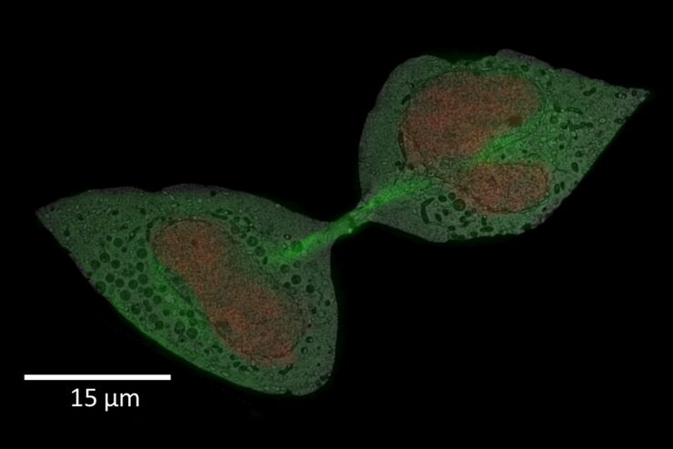
The live-cell CLEM workflow allows you to capture dynamic information related to a relevant biological process as it happens and put these observations into their ultrastructural context. The Leica…
Loading...
Advancing Cellular Ultrastructure Research
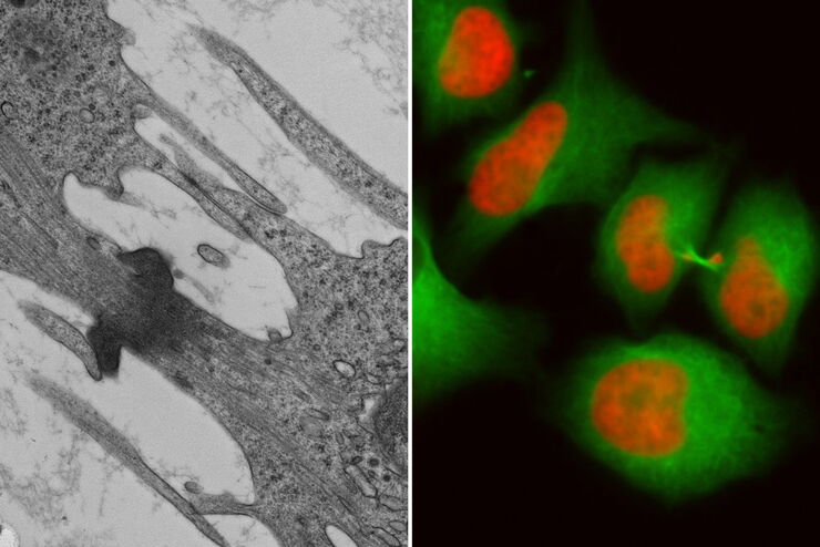
Freeze-fracture and freeze-etching are useful tools for studying flexible membrane-associated structures such as tight junctions or the enteric glycocalyx. Freeze-fracture and etching are two…
Loading...
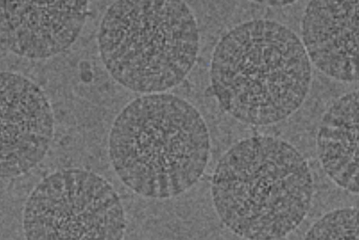
冷冻光电联用(Cryo-CLEM)之旅
本文主要介绍Cryo-CLEM技术及其为科学家带来的便益。此外,还特别说明了一些相关文献。
近期在冷冻电子显微镜工作流程领域取得的技术进步,让我们能够获取到细胞蛋白质社会学的3D数据,其分辨率更是达到前所未有的1纳米以下。工作流程中有一个步骤,需要从样品获取目标位置纳米级分辨率的图像,而要得到这样的结果,就需要用到冷冻光学显微镜。这种显微镜如果用于低温电子显微镜工作流程,通常就称为Cryo…
Loading...
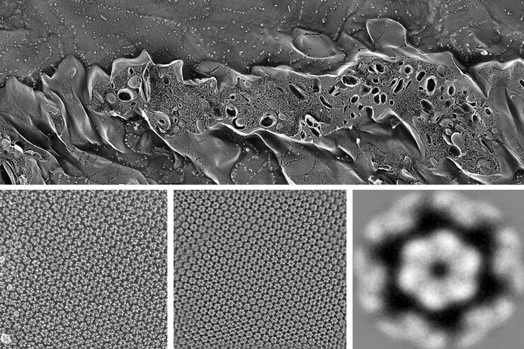
![[Translate to chinese:] 3D reconstruction of an intercellular bridge in a C. elegans embryo](/fileadmin/_processed_/0/9/csm_cryo-workflow-lp-mz_d30fabea6b.jpg "[Translate to chinese:] 3D reconstruction of an intercellular bridge in a C. elegans embryo")
下载 EM 工作流程解决方案手册
这本 26 页的工作流程手册中讨论的解决方案已被证明能够提供快速、可靠和可重复的结果。如果您对自己的工作流程有特殊要求,或对此处显示的主题有任何疑问,我们的徕卡专家将随时乐意为您提供帮助。
Loading...
Exploring the Structure and Life Cycle of Viruses
The SARS-CoV-2 outbreak started in late December 2019 and has since reached a global pandemic, leading to a worldwide battle against COVID-19. The ever-evolving electron microscopy methods offer a…
Loading...
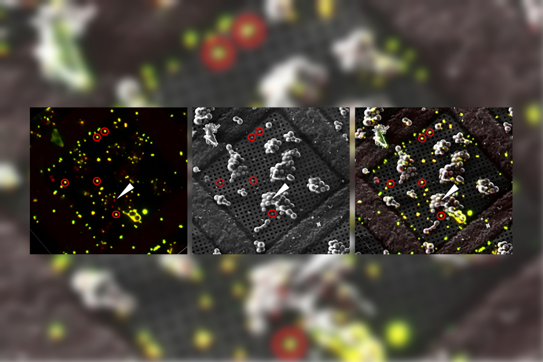
![[Translate to chinese:] Cryo FIB lamella - Overlay of SEM and confocal fluorescence image. Target structure in yeast cells (nuclear pore proteine Nup159-Atg8-split Venus, red) marked by an arrow. Scale bar: 5 µm. Alegretti et al., Nature 586, 796-800 (2020).](/fileadmin/_processed_/c/d/csm_Targeting_Nuclear_Pore_Complexes_teaser_16478dc18a.jpg "[Translate to chinese:] Cryo FIB lamella - Overlay of SEM and confocal fluorescence image")
使用冷冻共聚焦显微镜定位活性循环核孔复合物
本文介绍了如何利用冷冻光学显微镜,尤其是冷冻共焦显微镜来提高冷冻工作流程的可靠性。评估了EM网格和样品的质量,并分析了目标结构的分布。本文展示了如何将冷冻共焦3D数据投射到SEM图像上,将感兴趣结构可靠地保留在FIB切割的薄片内,以便在冷冻TEM中进行进一步研究。
Loading...
Advancing Cell Biology with Cryo-Correlative Microscopy
Correlative light and electron microscopy (CLEM) advances biological discoveries by merging different microscopes and imaging modalities to study systems in 4D. Combining fluorescence microscopy with…
Loading...
Workflows and Instrumentation for Cryo-electron Microscopy
Cryo-electron microscopy is an increasingly popular modality to study the structures of macromolecular complexes and has enabled numerous new insights in cell biology. In recent years, cryo-electron…