Loading...

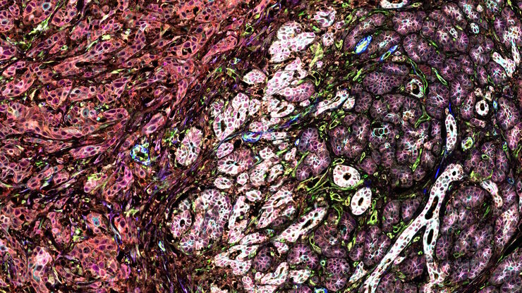
IBEX, Cell DIVE, and RNA-Seq: A Multi-omics Approach to Follicular Lymphoma
In a recent study by Radtke et al., a multi-omics spatial biology approach helps shed light on early relapsing lymphoma patients
Loading...
![[Translate to chinese:] 40x magnification of organoids cluster taken on Mateo TL.Cell type: esophageal squamous carcinoma; scale bar 15µm. Courtesy of bioGenous, China. [Translate to chinese:] 40x magnification of organoids cluster taken on Mateo TL.Cell type: esophageal squamous carcinoma; scale bar 15µm. Courtesy of bioGenous, China.](/fileadmin/_processed_/c/a/csm_Esophageal_squamous_carcinoma_scale__bar_15_m_7bfa184294.jpg)
克服类器官三维细胞培养中的观察挑战
类器官在细胞生物学和药物发现中至关重要,因为它们能够模拟体内细胞的复杂性和结构,有助于癌症等微环境至关重要的疾病研究。类器官可根据患者的基因型进行定制,这也有助于个性化医学研究。
Loading...
![[Translate to chinese:] Image of burrs (red arrows) at the edge of a battery electrode acquired with a DVM6 digital microscope. [Translate to chinese:] Image of burrs (red arrows) at the edge of a battery electrode acquired with a DVM6 digital microscope.](/fileadmin/_processed_/7/6/csm_Burrs_at_the_edge_of_a_battery_electrode_effd6b277b.jpg)
电池制造过程中的毛刺检测
毛刺是电池电极片边缘可能出现的缺陷,例如在制造过程中的分切环节。它们可能会因诸如短路等故障导致电池性能下降,并引发安全和可靠性问题。毛刺检测是电池生产质量控制的重要部分,对于生产具有可靠性能和寿命的电池至关重要。通过适当照明的光学显微镜可以在生产过程的关键步骤中快速可靠地对电极上的毛刺进行视觉检测。
Loading...

How do Cells Talk to Each Other During Neurodevelopment?
Professor Silvia Capello presents her group’s research on cellular crosstalk in neurodevelopmental disorders, using models such as cerebral organoids and assembloids.
Loading...
![[Translate to chinese:] Camera image during auto alignment. The feedback lines indicate if the correct edges in the image are detected. Green: Vertical center line [Translate to chinese:] Camera image during auto alignment. The feedback lines indicate if the correct edges in the image are detected. Green: Vertical center line; Magenta: Upper edge of the light gap; White: Lower edge of the light gap (not visible here, falling together with red line); Red: Knife edge; Blue: Left and right edge of the block face being automatically detected.](/fileadmin/_processed_/d/1/csm_Camera_image_during_auto_alignment_UC_Enuity_c148ecb865.jpg)
高质量超薄切片:样品与切片刀自动对齐
超薄切片技术是获取样品切片的最常用方法。在室温条件制备时,将样品小块嵌入环氧树脂中,然后通过修剪去除多余的树脂,并使用玻璃刀或金刚石刀将样品切成厚度为50-100纳米之间的薄片。
Loading...
![[Translate to chinese:] Multicolor fixed STED image. Inner ear section, mouse, TauSTED Xtend 589 on AF488 and TauSTED Xtend 775 on AF633-Phalloidin. [Translate to chinese:] Multicolor fixed STED image. Inner ear section, mouse, TauSTED Xtend 589 on AF488 and TauSTED Xtend 775 on AF633-Phalloidin. Sample courtesy of Dennis Derstrof, Klinik für Hals-, Nasen und Ohrenheilkunde, Universität Marburg & Prof. Dr. Dominik Oliver aus dem Institut für Physiologie und Pathophysiologie, Abteilung für Neurophysiologie, Universität Marburg.](/fileadmin/_processed_/c/a/csm_Inner_ear_section_mouse_multicolor_fixed_TauSTED_Xtend_1a1a0b424b.jpg)
细胞活成像的纳米级扩展
新的STED显微技术方法——TauSTED Xtend,使得在纳米级别下对活体完整样本进行扩展多色成像成为可能。通过结合空间和寿命信息,TauSTED Xtend提供了额外一层信息,允许在极低的光剂量下分辨小细节并在整体结构中解析它们。
Loading...
![[Translate to chinese:] Multicolor TauSTED Xtend 775 for Cell Biology applications that require nanoscopy resolution for multiple cellular components. Cells showing vimentin fibrils (AF 594), actin network (ATTO 647N), and nuclear pore basket (CF 680R). [Translate to chinese:] Multicolor TauSTED Xtend 775 for Cell Biology applications that require nanoscopy resolution for multiple cellular components. Cells showing vimentin fibrils (AF 594), actin network (ATTO 647N), and nuclear pore basket (CF 680R). Sample courtesy of Brigitte Bergner, Mariano Gonzales Pisfil, Steffen Dietzel, Core Facility Bioimaging, Biomedical Center, Ludwig-Maximilians-University, Munich, Germany.](/fileadmin/_processed_/5/4/csm_Triple_color_fixed_sample_TauSTED_Xtend_2_3_5d4d9caa21.jpg)
STED样品制备指南
这份指南旨在帮助用户优化受激发射损耗(STED)纳米成像的样品制备,特别是在使用徕卡微系统的STED显微镜时。它提供了单色STED成像用荧光标记的概述,并对其性能进行了评级。

![[Translate to chinese:] THUNDER image of brain-capillary endothelial-like cells derived from human iPSCs (induced pluripotent stem cells) where cyan indicates nuclei and magenta tight junctions. [Translate to chinese:] THUNDER image of brain-capillary endothelial-like cells derived from human iPSCs (induced pluripotent stem cells) where cyan indicates nuclei and magenta tight junctions.](/fileadmin/_processed_/8/e/csm_Brain-capillary_endothelial-like_cells_derived_from_human_iPSCs_b5dcf04bc9.jpg)
![[Translate to chinese:] Section ribbons with increasing section thickness - silver to purple ending in blue sections. [Translate to chinese:] Section ribbons with increasing section thickness - silver to purple ending in blue sections.](/fileadmin/_processed_/7/2/csm_Section_ribbons_with_increasing_section_thickness_8b0ba46e79.jpg)