
Loading...

Gene Editing with CRISPR/Cas9 - Breakthrough in Genome Engineering
The CRISPR/Cas9 system is one of several different bacterial systems for defense against viral attacks. It consists of two main components. One is a small piece of RNA which binds to the viral target…
Loading...
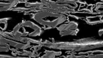
Paper Samples - Sample Preparation for SEM
Application Note for Leica EM RES102 - A coated paper sample has been prepared with ion beam slope cutting in order to test the procedure with regard to its applicability. With the use of ion beam…
Loading...

Imaging and Analyzing Zebrafish, Medaka, and Xenopus
Discover how to image and analyze zebrafish, medaka, and Xenopus frog model organisms efficiently with a microscope for developmental biology applications from this article.
Loading...
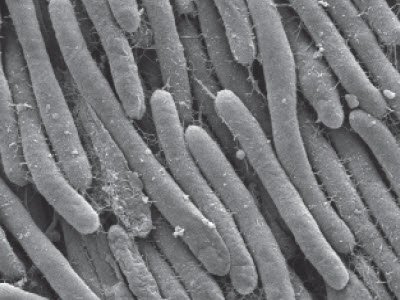
细菌实验方案 - 对大肠杆菌样本实施临界点干燥以进行SEM分析
徕卡EM CPD300的应用文档 - 生命科学研究,对大肠杆菌样本实施临界点干燥后,进行铂/钯金属镀膜,后用SEM拍摄微观形貌,将样本放入一个滤盘中(孔径:16-40μm),置入滤盘和多孔样品架内。在含有培养基的琼脂上培养真菌和细菌,为期3天。选取部分细菌菌落。
Loading...
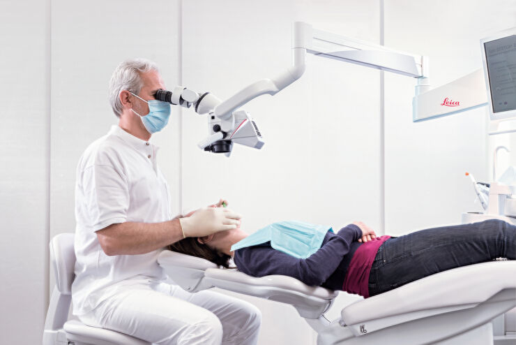
使用牙科手术显微镜成功进行牙髓治疗
在牙髓治疗中,准确的治疗不仅取决于牙医的技术技能和知识,还取决于手术区域清晰、详细的可视化。由于牙髓治疗的结果受到许多肉眼无法看到的因素的影响,例如额外的根管或解剖变异,牙科手术显微镜提供的高放大倍率和照明已成为诊断和治疗不可或缺的工具。如今,人们普遍认为牙科手术显微镜的使用有助于扩大牙髓治疗的潜力。
Loading...

Investigating Fruit Flies (Drosophila melanogaster)
Learn how to image and investigate Drosophila fruit fly model organisms efficiently with a microscope for developmental biology applications from this article.
Loading...

Studying Caenorhabditis elegans (C. elegans)
Find out how you can image and study C. elegans roundworm model organisms efficiently with a microscope for developmental biology applications from this article.
Loading...
BABB Clearing and Imaging for High Resolution Confocal Microscopy
Multipohoton microscopy experiment using Leica TCS SP8 MP and Leica 20x/0.95 NA BABB immersion objective.
Understanding kidney microanatomy is key to detecting and identifying early events in kidney…
Loading...
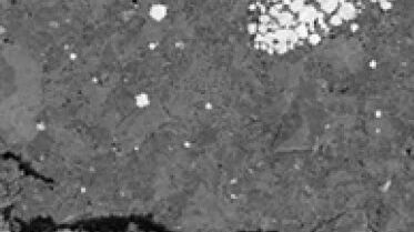
Ion Beam Polishing of Sample Surfaces - Sample Preparation for SEM
Application Note for Leica EM RES102 - Ion milling can be used to reduce the roughness of sample surfaces. Small angles less than 6° with respect to the sample surface are necessary. The high voltage…