Leica M165 FC
体视显微镜
复合光学显微镜
产品
首页
Leica Microsystems
Leica M165 FC 荧光体视显微镜
阅读我们的最新文章
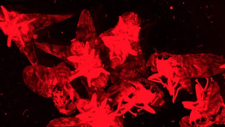
Investigating Fruit Flies (Drosophila melanogaster)
Learn how to image and investigate Drosophila fruit fly model organisms efficiently with a microscope for developmental biology applications from this article.
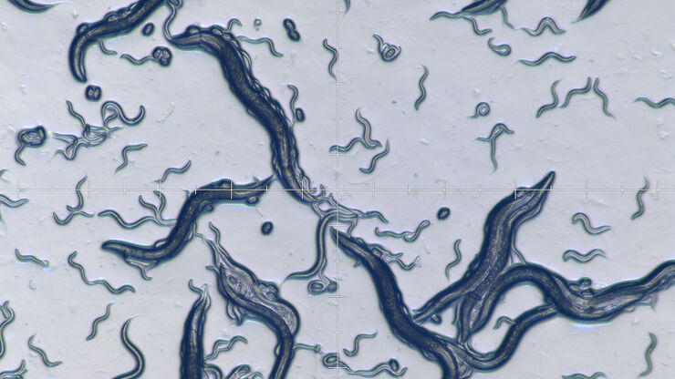
Studying Caenorhabditis elegans (C. elegans)
Find out how you can image and study C. elegans roundworm model organisms efficiently with a microscope for developmental biology applications from this article.
体视显微镜的历史
本文概述了从 1600 年至今体视显微镜的发展和演变。直到 19 世纪中叶,所有光学显微镜都是手工制作的。由于无法准确预测透镜的特性,因此必须通过反复试验来制作和测试透镜,直到达到理想的效果。
想了解更多信息?
请咨询我们的专家。
您想获取专人咨询吗? Show local contacts


![[Translate to chinese:] A portion of an early binocular microscope developed by John Leonhard Riddel in the early 1850s. [Translate to chinese:] A portion of an early binocular microscope developed by John Leonhard Riddel in the early 1850s.](/fileadmin/_processed_/1/8/csm_Early_binocular_microscope_by_John_Leonhard_Riddel_teaser_60cc11264a.jpg)
